Total hip arthroplasty: Techniques and results
Primary total hip arthroplasty has become one of the most successful surgical procedures over the past 50 years and is currently performed worldwide with similar techniques and excellent results. Despite variations in technique and implant selection, medium and long-term outcome studies have demonstrated over 90% implant survival at 15 to 20 years. Previous problems with implant fixation have now been reduced, and the focus has shifted to the selection of improved bearing surfaces to limit wear, hip replacement options for younger patients, and improved surgical and anesthetic techniques. Current surgical approaches to the hip rely most often on direct lateral or posterolateral exposure. The most commonly utilized bearing surface for both hip replacement and hip resurfacing in Canada is a metal (cobalt-chrome) femoral head combined with a second-generation cross-linked polyethylene, combined with cementless implant fixation. Alternative bearings such as ceramic-on-ceramic and metal-on-metal may be considered for hip replacement in younger patients. Although it has not been determined which surface will prove best for younger patients in the long-term, there is no question about the benefits of total hip arthroplasty. With current techniques, the results are favorable, and patient satisfaction, pain relief, and long-term implant survival are excellent.
Younger, more active patients are now candidates for total hip replacement with the advent of improved implant fixation and new low-wearing bearing surfaces.
The current long-term success of total hip replacement (THR) surgery has led to the observation by Coventry[1] that “total hip replacement, indeed, might be the orthopaedic operation of the century.”
The indications for THR have expanded to such an extent that this surgery is no longer performed only in the elderly or in those with debilitating hip pain, arthritis, and severe functional restrictions.
Rather, THR is now performed in younger and higher-demand patients, with expectations, quality-of-life measures, and intentions to return to prior activity levels that challenge surgical techniques and implant design technology.
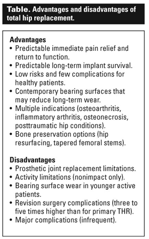
The advantages of THR generally outweigh the disadvantages (Table), and attention is now focused on improved fixation of the implants, reduction in the rates of failure, and development of bearing surfaces to reduce long-term wear and improve implant longevity.
Surgical exposure
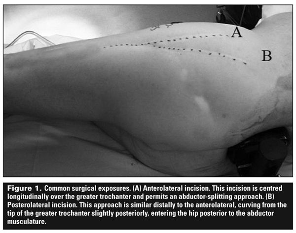
Several surgical exposures are utilized for THR. The two most common exposures (Figure 1) are the anterolateral[2] and the posterolateral approaches to the hip.[3]
Patients may also be offered one of the newer techniques of surgical exposure referred to as muscle-sparing or minimally invasive.
The decision of which surgical exposure to use will depend upon surgeon experience and preference, patient body habitus (i.e., obesity), patient anatomical factors, the location and type of prior surgical incisions over the hip, and implant selection. The most important factor to consider is surgeon experience and preference.
The anterolateral exposure is an abductor-splitting approach requiring removal and repair of the anterior 30% to 40% of the gluteus medius and minimus. This approach may also be utilized for revision THR surgery.
Many surgeons select this approach based upon the potential for a reduced dislocation rate. Disadvantages of the anterolateral approach include:
• An increase in limp due to splitting of the abductor muscle (also likely due to traction injury to anterior branches of the superior gluteal nerve during surgery). Often the limp is reported as being asymptomatic, but frequently it is a Trendelenburg gait.
• An increase in the formation of heterotopic bone within the abductor muscles and anteriorly over the capsule and greater trochanter.
• A greater incidence of trochanteric complications (intraoperative fracture, postoperative fracture, or escape of the greater trochanter), and trochanteric pain (often incorrectly attributed to a diagnosis of trochanteric bursitis), most likely due to failure of the abductors to heal following the repair.
• A tendency for the surgeon to insert the femoral component angled from anterior to posterior within the femoral canal (i.e., nonanatomic femoral component placement).
With the popularity of less invasive surgery, the posterolateral exposure has again gained prominence. Disadvantages of the posterolateral approach include:
• Perhaps a slightly higher risk of dislocation, although with experience this is minimized.
• The need for careful attention to component orientation in order to insert the implants in proper anteversion.
In Canada between 2008 and 2009, the direct lateral approach (60%) and posterolateral approach (36%) combined for over 95% of all surgical exposures.[4]
When minimally invasive surgery for THR is performed, it is most commonly performed using one of these two approaches. Other minimally invasive surgical approach options include the two-incision approach,[5,6] the anterolateral (Watson-Jones) approach, and the direct anterior (Hueter) approach.[7]
Often these surgical approaches require the surgeon to change to a different OR setup[6] (i.e., one with a specialized table, retractors, and lights, and access to intraoperative X-ray) and to use an implant he or she may be less familiar with in order to make the procedure feasible.
While there may be a few short-term advantages to minimally invasive surgery, the early and mid-term results have been associated with significantly increased risks and surgical complications,[5] which have not been seen in THR prior to the popularity of these techniques.
Thus, the enthusiasm for minimally invasive surgery has declined recently in favor of surgery performed safely through smaller incisions, and with the goal of achieving an ideal implant orientation and longevity.
Computer-assisted surgery (CAS) for total hip replacement has gained popularity and is performed in many centres. The advantages and results of CAS have been difficult to assess, and there does not appear to be any significant advantage to CAS at this time.
The one area of potential advantage is that CAS may be useful in identifying “outlier” acetabular component position/angulation and leg length and hip offset intraoperatively, which might help in select situations, especially for surgeons with less experience performing THR and surgeons combining CAS with minimally invasive surgery.
The main disadvantage is increased OR time and increased cost. Overall, CAS has not been shown to be cost-effective to date.
Implant fixation: Cemented or cementless?
Both cemented and cementless fixation are currently utilized in THR surgery, although there has been a trend in North America toward cementless implants over the past 10 years.
Total hip replacement implants typically consist of the acetabular component (which is fitted into the patient’s native acetabular pelvic bone with or without cement), the femoral component (inserted down the femoral canal), and the bearing surfaces (the articulating aspects of the implant).
When describing fixation methods, we are referring to the femoral and acetabular components.
Acetabular component implant fixation
The use of cemented acetabular components has declined in recent years in North America, although cemented components are still used occasionally in older and lower-demand patients.
When compared with cementless implants, cemented acetabular components have been associated with increased rates of loosening at 10 to 20 years, especially in patients younger than 50,[8] when compared to cementless implants.
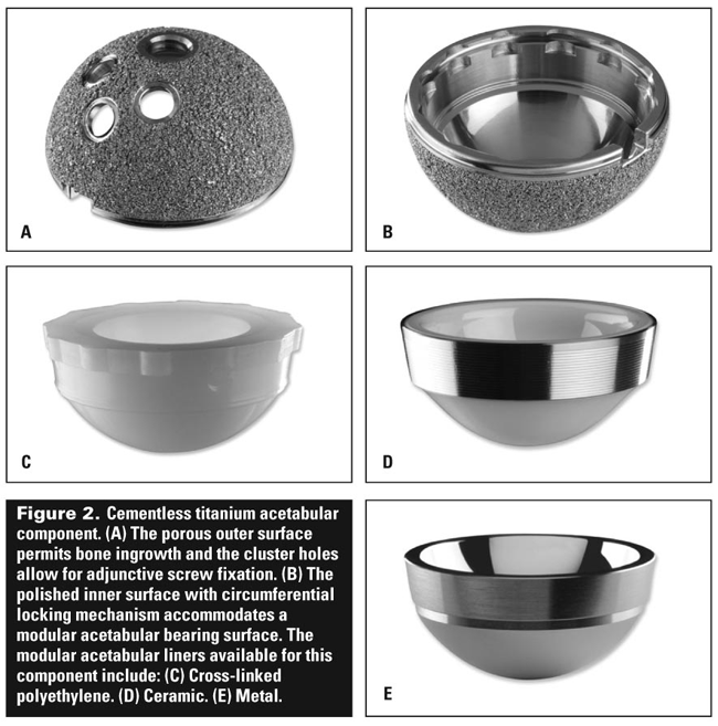
Cementless acetabular fixation was introduced to solve the problem of loosening with cemented acetabular cups. The most commonly used composite for cementless acetabular components is titanium alloy, which is favorable for bone ingrowth.
Typically, a modular bearing surface (the liner) is inserted into the inner aspect of the acetabular component, and locks into place via a mechanism contained within the acetabular component.
The acetabular component may accept bearing surfaces, including liners made of polyethylene, ceramic, or metal, to complete the acetabular component composition (Figure 2).
This modular bearing surface may be exchanged in the future if wear or other less common indications make this necessary, leaving the intact osseo-integrated acetabular component in place.
The long-term results of cementless titanium acetabular fixation have been favorable. At a minimum of 20 years, the implant survival for titanium hemispherical cups has recently been reported at over 95%.[9]
However, wear-related complications of the polyethylene liner inside and on the backside (and of the associated modular locking mechanism) occur in approximately 20% of patients by 20 years, a problem that has become the focus of research in THR surgery.
Femoral component implant fixation
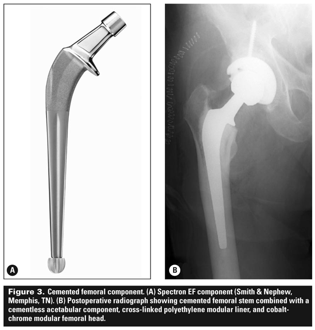
Cemented femoral component fixation has achieved excellent long-term results in multiple studies at 17 to 30 years[10-14] and continues to be the gold standard against which the more popular cementless femoral fixation must be measured.
Contemporary cementing techniques were refined in the 1970s and require attention to detail. In addition to cement technique, there are two implant designs: the cemented tapered polished collarless stem (Exeter, Stryker Orthopaedics, Mahwah NJ) and the Spectron EF stem (Smith & Nephew Orthopaedics, Memphis TN) (Figure 3) which have incorporated differing design characteristics, yet which have both proven very successful in the long-term clinical trials.[15,16]
Early failures of cemented stems implanted with older cementing technique included loosening, stem fracture, and localized areas of bone destruction (osteolysis) from cement wear debris. Cementless implants were developed to solve these problems.
Today, cementless femoral components are produced in various designs and shapes, and with different metallic compositions and surface preparation to promote osseo-integration.
All uncemented femoral stem designs rely on metaphyseal fixation, metaphyseal-diaphyseal junction fixation, diaphyseal fixation, or a combination of the three.
The tapered titanium alloy cementless stem (Figure 4) has grown in popularity[17] and is becoming commonly used worldwide. Achieving a press-fit via a single or dual tapered wedge with subsequent proximal osseo-integration of bone has proven successful in multiple long-term studies[18] of tapered titanium stems, with over 95% survival at 10 to 20 years.
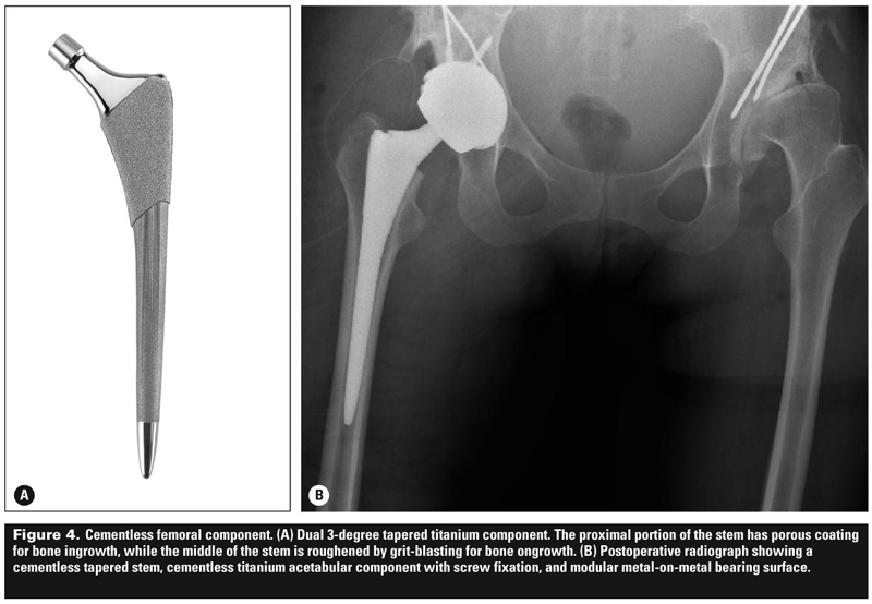
In summary, while cemented femoral stem fixation remains the gold standard in long-term studies, it is highly dependent on cementing technique and implant design.
Cemented acetabular fixation is rarely utilized in North America. Cementless fixation on both the femoral and acetabular sides is performed most commonly and relies on an immediate press-fit of the implant followed by osseo-integration into host bone.
Hip resurfacing
Total hip resurfacing, also known as surface replacement arthroplasty or hip resurfacing (HR), has gained in popularity partly because of two metal-on-metal HR implants approved by the FDA within the past 9 years.
HR has been performed for 15 years in both North America and Europe with favorable results.[19,20] It is performed using a cemented metal femoral component shaped to the patient’s native femoral head and a cementless acetabular component with a polished inner cobalt-chrome metal surface (Figure 5).
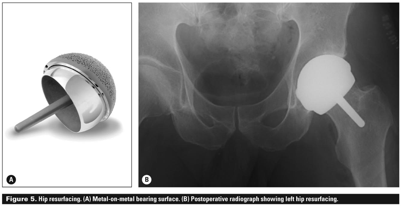
The two surfaces join to create a metal-on-metal bearing surface that has low-wear properties. Relative indications for HR surgery[21] include younger age, active occupational and lifestyle requirements, favorable bone anatomy and quality (without cystic change, defects, or dysplasia), normal weight, and male sex.
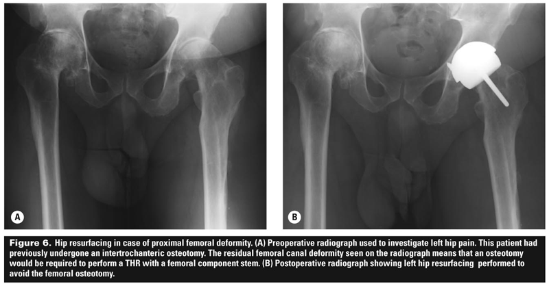
Hip resurfacing may also be used adjunct when there is proximal femoral deformity that would otherwise require an osteotomy to perform a THR (Figure 6).
Contraindications include impaired renal function (or the potential for impairment with a diagnosis such as diabetes) with an inability to process serum metal ions, older age, osteoporosis or osteopenia, unfavorable femoral head geometry, clinical metal sensitivity history (usually a nickel sensitivity), a leg-length discrepancy greater than 1 cm, and women of childbearing age.
The primary concern regarding HR in younger women is how the increased ion levels of cobalt and chromium normally associated with a metal-on-metal bearings could effect fetal development, as these ions do cross the placenta.
Two recent studies suggest that although these ions cross the placenta, a modulatory effect occurs, decreasing their concentration in the fetus. Still, such results should be interpreted with caution.[22,23]
Hip resurfacing surgery is performed with similar exposures to those used in conventional THR. Contrary to popular belief, hip resurfacing is not a minimally invasive procedure.
Rather, it often requires a larger incision and surgical exposure, with additional soft tissue capsular releases that are not typically performed in THR—thus HR is often more invasive, not less.
Despite this, recovery following hip resurfacing is similar to conventional THR, likely due to generally younger patient age. The proposed advantages (which remain controversial) of HR surgery include:
• Bone preservation on the femoral side.
• Ease of future revision surgery on the femoral side.
• Large-head bearing surface with a reduced dislocation rate.
• Use of a metal-on-metal low-wear bearing surface.
• Patient findings that HR feels more normal than THR.
These advantages, however, can all be obtained from conventional THR with the use of a metal-on-metal bearing surface, particularly if a large femoral head is used.
Surgeons who disfavor hip resurfacing do so for several reasons:
• Bone preservation may not necessarily occur, with occasionally more bone being removed on the acetabular side to achieve a deepened socket with a press-fit and no option for screw fixation.
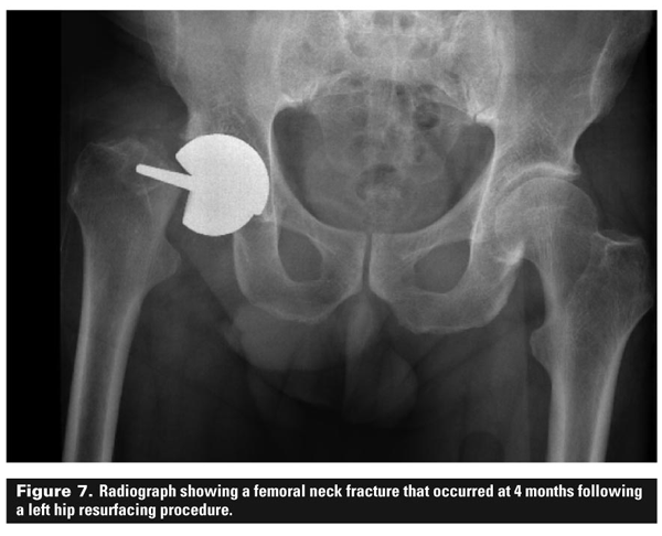
• The risk of notching the femoral neck and subsequent femoral neck fracture (risk 0.8%–1.5%)[24,25] (Figure 7).
• Elevated levels of serum and urine cobalt, chromium, molybdenum, and selenium ions that remain elevated lifelong.
• The risk of lymphocyte-mediated metal sensitivity reactions and/or the development of pseudotumors, recently highlighted in research at UBC and McGill University.[26]
• It is a technically more demanding surgical procedure for the surgeon and team, with a steep learning curve[27] and potentially increased risks and complications when compared with conventional THR.
While HR is an option to consider in younger and more active patients, it requires careful preoperative assessment and a discussion with the patient about all of the issues, including the risk of increased metal ion levels and metal sensitivity reactions, and the low risk of psuedotumor.[28]
In addition, impact activities are not encouraged after HR, and the restrictions and precautions following surgery are similar to those for THR. Overall, the short-term results of HR (up to 5 years) have been worse than for THR, and therefore hip resurfacing should be used with caution. THR remains the gold standard.
Bearing surfaces
With current implant fixation methods demonstrating excellent long-term results, the bearing surface in THR is now the focus of much research.
The bearing surface is where the movement of the two bearings occurs and which provides the range of motion and articulation of the prosthetic ball and socket joint.
Within the last 10 years, the use of traditional ultrahigh molecular weight polyethylene (UHMWPE) acetabular liners has declined with the development of new kinds of polyethylene.
Highly cross-linked polyethylenes
To reduce wear rates and particulate debris, highly cross-linked polyethylene (XLPE) has been used in total hip arthroplasty for 8 years.
The manufacturing process for these materials cross-links the molecules and improves wear characteristics but slightly reduces the strength of the polyethylene.
Free radicals may be generated in the process, potentially allowing for oxidative changes in the polyethylene, unless these changes are appropriately managed in the manufacturing process.
Thus, the ideal XLPE would be cross-linked at an appropriate level of radiation, and then remelted to remove these free radicals and thus reduce the oxidation process. Currently, all of the THR implant manufacturers produce either a first-generation or second-generation XLPE.
When combined with a polished cobalt-chrome head of multiple sizes, these new XLPEs have shown promise in reducing in vivo and simulator wear measurements significantly[29] compared with traditional UHMWPE.
The increase in wear resistance is, however, associated with a decrease in fatigue strength and toughness. The use of XLPE liners requires meticulous positioning of the acetabular component to avoid vertical placement of the implant, which reports have associated with an increased risk of fracture at the rim of the polyethylene liner (Figure 8).
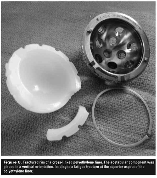
The use of XLPE has allowed the introduction of larger femoral heads, which increase the stability of the hip with their greater diameter and increased “jump distance.”
When XLPE is used, wear rates of the polyethylene have not been shown to be worse with larger femoral heads. This is in contrast to older UHMWPE, which demonstrates higher volumetric polyethylene wear as the size of the femoral head is increased.
Alternative bearing surfaces
Other bearing surfaces have been developed and utilized in THR in an attempt to reduce the wear-related polyethylene complications.
Polyethylene wear and debris formation result in hip joint synovitis, joint instability, osteolysis, and, potentially, prosthesis loosening.
Alternative bearing surfaces such as metal-on-metal, ceramic-on-ceramic, ceramic-on-XLPE, oxinium (oxidized zirconium), and even the new XLPEs themselves have been developed in an attempt to reduce wear and improve implant survival in younger and more active patients.
Currently in Canada, the most commonly utilized bearing surface is a cobalt-chrome head combined with cross-linked polyethylene (59%), while other alternative bearings such as metal-on-metal (11% ; includes HR use) and ceramics (13%) are used less frequently, and usually in younger patients.[4]
Ceramics. Alumina ceramics were introduced in the 1970s. They have a very low coefficient of friction and demonstrate the lowest wear rates of any implant bearing surface.[30] They are scratch resistant and may be combined as a modular ceramic acetabular liner with a ceramic head.
There is no potential for metal ion release, which is attractive to younger patients, especially females of childbearing age. Although ceramics can fracture because of their brittle composition, the rate of fracture is very low (0.5%)[31] in most studies.
Newer ceramic composites of alumina (Biolox Delta Ceramic, CeramTec AG, Lauf, Germany) have demonstrated increased strength and fracture resistance, and offer increased neck-length options intraoperatively (Figure 9).
Ceramic-on-ceramic bearing surfaces have been associated with squeaking that is audible to the patient and others.
Initially believed to occur rarely (~1%) in ceramic-on-ceramic THR, recent studies have shown that noise (squeaking, grinding, rubbing, or other audible sounds from the hip) occurs more frequently than originally reported, and is experienced by 10% to 17% of patients with a ceramic-on-ceramic bearing surface.[32,33]
The causes and implications of squeaking have yet to be determined, but are likely to be multifactorial: acetabular modular implant design-specific factors, component orientation and malposition, instability, and femoral component design have all been implicated.
The use of ceramic-on-ceramic bearings offers many advantages in terms of wear reduction, especially for young and active patients.
Nonetheless, patients considering ceramic-on-ceramic bearings should be informed of this phenomenon, and the surgeon and patient should discuss avoiding ceramic implants associated with a higher incidence of squeaking. There are no long-term clinical results to date for the newer ceramic composites.
Oxinium. Oxidized zirconium (Smith & Nephew, Memphis, TN) has been developed for femoral head components and has the wear-resistance of ceramic without the brittle fracture risk.
Compared with the limited ceramic ball neck lengths available, oxinium allows for increased length options intraoperatively. No long-term clinical studies of this material have been published yet, and it is only available from one manufacturer.
Metal-on-metal. Metal-on-metal bearing surfaces have been used widely since the 1960s.[34-36] Poor metallurgy, poor design (equatorial head edge bearing), and poor fixation led to early failures of many hip replacements using metal bearings.
However, a subset of these were found to have a suitable central-head bearing and minimal wear when compared with hip replacements using UHMWPE.
This finding led to a resurgence of interest in metal-on-metal surface bearings, and an attempt to create a bearing surface with similar metallurgy and design to that found in the subset with long-term survival.
Metal bearing surfaces demonstrate very low wear rates—somewhere between rates for ceramic-on-ceramic and metal-on-XLPE—and much less wear than for conventional UHWMPE.
Metal bearings support the use of a larger femoral head size, which demonstrates better fluid-film lubrication, and lower metal ion levels than found with smaller head combinations, making metal-on-metal ideally suited for hip resurfacing.
Metal is not brittle like ceramic, making it attractive for younger patients. Larger head sizes are also associated with improved joint stability and a reduced risk of dislocation.
While metal-on-metal bearing surfaces generally are associated with elevated metal ion levels,[37] no long-term effects are known.
Preoperatively, patients must be informed that the low risk of metal sensitivity and lymphocyte-mediated reaction is similar to that for hip resurfacing.
Recently, inflammatory granulomatous pseudotumors, which are necrotic cystic soft tissue tumors, have been seen following large-head metal-on-metal hip replacement with one or more implant designs, and have been seen less often following HR.
For this reason, metal-on-metal bearing surfaces should be used with caution in THR, patients should be followed closely at yearly intervals, and patients should be counseled about the possibility of metal-related complications that will lead to poor outcome if they occur, even after revision surgery.
Conclusions
Total hip arthroplasty has become the treatment of choice for hip-related disorders leading to arthritis in the adult population. With improvements in long-term clinical results, implant fixation, and new low-wear bearing surfaces, THR surgery is now being performed in younger and more active patients.
Using current implant design and techniques, the implant survival at 20 years is favorable, with over 90% implant survival in multiple studies.
However, with younger and more active patients undergoing total hip replacement, the challenge will be the bearing surface selection. It remains to be determined which bearing surfaces will provide the lowest wear rates and the fewest wear-related complications in the long term.
Competing interests
None declared.
References
![]() 1. Coventry MB. Foreword. In: Amstutz HC (ed). Hip arthroplasty. New York: Churchill Livingstone; 1991.
1. Coventry MB. Foreword. In: Amstutz HC (ed). Hip arthroplasty. New York: Churchill Livingstone; 1991.
![]() 2. Mulliken BD, Rorabeck CH, Bourne RB, et al. A modified direct lateral approach in total hip arthroplasty: A comprehensive review. J Arthroplasty 1998;13:737-747.
2. Mulliken BD, Rorabeck CH, Bourne RB, et al. A modified direct lateral approach in total hip arthroplasty: A comprehensive review. J Arthroplasty 1998;13:737-747.
![]() 3. Kwon MS, Kuskowski M, Mulhall KJ, et al. Does surgical approach affect total hip arthroplasty dislocation rates? Clin Orthop Relat Res 2006;447:34-38.
3. Kwon MS, Kuskowski M, Mulhall KJ, et al. Does surgical approach affect total hip arthroplasty dislocation rates? Clin Orthop Relat Res 2006;447:34-38.
![]() 4. Canadian Institute for Health Information. Hip and knee replacements in Canada—Canadian Joint Replacement Registry (CJRR) 2008–2009 annual report. http://secure.cihi.ca/cihiweb/dispPage.jsp?cw_page=PG_1519_E&cw_topic=1519&cw_rel=AR_30_E (accessed 14 September 2010).
4. Canadian Institute for Health Information. Hip and knee replacements in Canada—Canadian Joint Replacement Registry (CJRR) 2008–2009 annual report. http://secure.cihi.ca/cihiweb/dispPage.jsp?cw_page=PG_1519_E&cw_topic=1519&cw_rel=AR_30_E (accessed 14 September 2010).
![]() 5. Bal BS, Haltom D, Aleto T, et al. Early complications of primary total hip replacement performed with a two-incision minimally invasive technique. Surgical technique. J Bone Joint Surg Am 2006;88:(suppl):221-233.
5. Bal BS, Haltom D, Aleto T, et al. Early complications of primary total hip replacement performed with a two-incision minimally invasive technique. Surgical technique. J Bone Joint Surg Am 2006;88:(suppl):221-233.
![]() 6. Berger RA, Duwelius PJ. The two-incision minimally invasive total hip arthroplasty: Technique and results. Orthop Clin North Am 2004;35:163-172.
6. Berger RA, Duwelius PJ. The two-incision minimally invasive total hip arthroplasty: Technique and results. Orthop Clin North Am 2004;35:163-172.
![]() 7. Seng BE, Berend KR, Ajluni AF, et al. Anterior-supine minimally invasive total hip arthroplasty: Defining the learning curve. Orthop Clin North Am 2009;40:343-350.
7. Seng BE, Berend KR, Ajluni AF, et al. Anterior-supine minimally invasive total hip arthroplasty: Defining the learning curve. Orthop Clin North Am 2009;40:343-350.
![]() 8. Barrack RL, Mulroy RD Jr, Harris WH. Improved cementing techniques and femoral component loosening in young patients with hip arthroplasty. A 12-year radiographic review. J Bone Joint Surg Br 1992;74:385-389.
8. Barrack RL, Mulroy RD Jr, Harris WH. Improved cementing techniques and femoral component loosening in young patients with hip arthroplasty. A 12-year radiographic review. J Bone Joint Surg Br 1992;74:385-389.
![]() 9. Della Valle CJ, Mesko NW, Quigley L, et al. Primary total hip arthroplasty with a porous-coated acetabular component. A concise follow-up, at a minimum of twenty years, of previous reports. J Bone Joint Surg Am 2009;91:1130-1135.
9. Della Valle CJ, Mesko NW, Quigley L, et al. Primary total hip arthroplasty with a porous-coated acetabular component. A concise follow-up, at a minimum of twenty years, of previous reports. J Bone Joint Surg Am 2009;91:1130-1135.
![]() 10. Ling RS, Charity J, Lee AJ, et al. The long-term results of the original Exeter polished cemented femoral component: A follow-up report. J Arthroplasty 2009;24:511-517.
10. Ling RS, Charity J, Lee AJ, et al. The long-term results of the original Exeter polished cemented femoral component: A follow-up report. J Arthroplasty 2009;24:511-517.
![]() 11. Herberts P, Malchau H. Long-term registration has improved the quality of hip replacement: A review of the Swedish THR Register comparing 160,000 cases. Acta Orthop Scand 2000;71:111-121.
11. Herberts P, Malchau H. Long-term registration has improved the quality of hip replacement: A review of the Swedish THR Register comparing 160,000 cases. Acta Orthop Scand 2000;71:111-121.
![]() 12. Mulroy RD Jr, Harris WH. The effect of improved cementing techniques on component loosening in total hip replacement. An 11-year radiographic review. J Bone Joint Surg Br 1990;72:757-760.
12. Mulroy RD Jr, Harris WH. The effect of improved cementing techniques on component loosening in total hip replacement. An 11-year radiographic review. J Bone Joint Surg Br 1990;72:757-760.
![]() 13. Issack PS, Botero HG, Hiebert RN, et al. Sixteen-year follow-up of the cemented spectron femoral stem for hip arthroplasty. J Arthroplasty 2003;18:925-930.
13. Issack PS, Botero HG, Hiebert RN, et al. Sixteen-year follow-up of the cemented spectron femoral stem for hip arthroplasty. J Arthroplasty 2003;18:925-930.
![]() 14. Carrington NC, Sierra RJ, Gie GA, et al. The Exeter Universal cemented femoral component at 15 to 17 years: An update on the first 325 hips. J Bone Joint Surg Br 2009;91:730-737.
14. Carrington NC, Sierra RJ, Gie GA, et al. The Exeter Universal cemented femoral component at 15 to 17 years: An update on the first 325 hips. J Bone Joint Surg Br 2009;91:730-737.
![]() 15. Williams HD, Browne G, Gie GA, et al. The Exeter Universal cemented femoral component at 8 to 12 years. A study of the first 325 hips. J Bone Joint Surg Br 2002;84:324-334.
15. Williams HD, Browne G, Gie GA, et al. The Exeter Universal cemented femoral component at 8 to 12 years. A study of the first 325 hips. J Bone Joint Surg Br 2002;84:324-334.
![]() 16. Garellick G, Malchau H, Herberts P. Survival of hip replacements. A comparison of a randomized trial and a registry. Clin Orthop Relat Res 2000;(375):157-167.
16. Garellick G, Malchau H, Herberts P. Survival of hip replacements. A comparison of a randomized trial and a registry. Clin Orthop Relat Res 2000;(375):157-167.
![]() 17. Danesh-Clough T, Bourne RB, Rorabeck CH, et al. The mid-term results of a dual offset uncemented stem for total hip arthroplasty. J Arthroplasty, 2007;22:195-203.
17. Danesh-Clough T, Bourne RB, Rorabeck CH, et al. The mid-term results of a dual offset uncemented stem for total hip arthroplasty. J Arthroplasty, 2007;22:195-203.
![]() 18. Lombardi AV Jr, Berend KR, Mallory TH, et al. Survivorship of 2000 tapered titanium porous plasma-sprayed femoral components. Clin Orthop Relat Res 2009;467:146-154.
18. Lombardi AV Jr, Berend KR, Mallory TH, et al. Survivorship of 2000 tapered titanium porous plasma-sprayed femoral components. Clin Orthop Relat Res 2009;467:146-154.
![]() 19. Treacy RB, McBryde CW, Pynsent PB. Birmingham hip resurfacing arthroplasty. A minimum follow-up of five years. J Bone Joint Surg Br 2005;87:167-170.
19. Treacy RB, McBryde CW, Pynsent PB. Birmingham hip resurfacing arthroplasty. A minimum follow-up of five years. J Bone Joint Surg Br 2005;87:167-170.
![]() 20. Amstutz HC, Le Duff MJ. Eleven years of experience with metal-on-metal hybrid hip resurfacing: A review of 1000 conserve plus. J Arthroplasty 2008;23(suppl):36-43.
20. Amstutz HC, Le Duff MJ. Eleven years of experience with metal-on-metal hybrid hip resurfacing: A review of 1000 conserve plus. J Arthroplasty 2008;23(suppl):36-43.
![]() 21. Della Valle CJ, Nunley RM, Barrack RL. When is the right time to resurface? Orthopedics 2008;31(suppl).
21. Della Valle CJ, Nunley RM, Barrack RL. When is the right time to resurface? Orthopedics 2008;31(suppl).
![]() 22. Ziaee H, Daniel J, Datta AK, et al. Transplacental transfer of cobalt and chromium in patients with metal-on-metal hip arthroplasty: A controlled study. J Bone Joint Surg Br 2007;89:301-305.
22. Ziaee H, Daniel J, Datta AK, et al. Transplacental transfer of cobalt and chromium in patients with metal-on-metal hip arthroplasty: A controlled study. J Bone Joint Surg Br 2007;89:301-305.
![]() 23. Amstutz HC, Antoniades JT, Le Duff MJ. Results of metal-on-metal hybrid hip resurfacing for Crowe type-I and II developmental dysplasia. J Bone Joint Surg Am 2007;89:339-346.
23. Amstutz HC, Antoniades JT, Le Duff MJ. Results of metal-on-metal hybrid hip resurfacing for Crowe type-I and II developmental dysplasia. J Bone Joint Surg Am 2007;89:339-346.
![]() 24. Shimmin AJ, Back D. Femoral neck fractures following Birmingham hip resurfacing: A national review of 50 cases. J Bone Joint Surg Br 2005;87:463-464.
24. Shimmin AJ, Back D. Femoral neck fractures following Birmingham hip resurfacing: A national review of 50 cases. J Bone Joint Surg Br 2005;87:463-464.
![]() 25. Amstutz HC, Campbell PA, Le Duff MJ. Fracture of the neck of the femur after surface arthroplasty of the hip. J Bone Joint Surg Am 2004;86-A:1874-1877.
25. Amstutz HC, Campbell PA, Le Duff MJ. Fracture of the neck of the femur after surface arthroplasty of the hip. J Bone Joint Surg Am 2004;86-A:1874-1877.
![]() 26. Garbuz DS, Tanzer M, Greidanus NV, et al. The John Charnley Award: Metal-on-metal hip resurfacing versus large-diameter head metal-on-metal total hip arthroplasty: A randomized clinical trial. Clin Orthop Relat Res 2009;468:318-325.
26. Garbuz DS, Tanzer M, Greidanus NV, et al. The John Charnley Award: Metal-on-metal hip resurfacing versus large-diameter head metal-on-metal total hip arthroplasty: A randomized clinical trial. Clin Orthop Relat Res 2009;468:318-325.
![]() 27. Nunley RM, Zhu J, Brooks PJ, et al. The learning curve for adopting hip resurfacing among hip specialists. Clin Orthop Relat Res 2009;468:382-391.
27. Nunley RM, Zhu J, Brooks PJ, et al. The learning curve for adopting hip resurfacing among hip specialists. Clin Orthop Relat Res 2009;468:382-391.
![]() 28. Counsell A, Heasley R, Arumilli B, et al. A groin mass caused by metal particle debris after hip resurfacing. Acta Orthop Belg 2008;74:870-874.
28. Counsell A, Heasley R, Arumilli B, et al. A groin mass caused by metal particle debris after hip resurfacing. Acta Orthop Belg 2008;74:870-874.
![]() 29. Bragdon CR, Kwon YM, Geller JA, et al. Minimum 6-year followup of highly cross-linked polyethylene in THA. Clin Orthop Relat Res 2007;465:122-127.
29. Bragdon CR, Kwon YM, Geller JA, et al. Minimum 6-year followup of highly cross-linked polyethylene in THA. Clin Orthop Relat Res 2007;465:122-127.
![]() 30. Semlitsch M, Willert HG. Clinical wear behaviour of ultra-high molecular weight polyethylene cups paired with metal and ceramic ball heads in comparison to metal-on-metal pairings of hip joint replacements. Proc Inst Mech Eng H 1997;211:73-88.
30. Semlitsch M, Willert HG. Clinical wear behaviour of ultra-high molecular weight polyethylene cups paired with metal and ceramic ball heads in comparison to metal-on-metal pairings of hip joint replacements. Proc Inst Mech Eng H 1997;211:73-88.
![]() 31. Capello WN, D’Antonio JA, Feinberg JR, et al. Ceramic-on-ceramic total hip arthroplasty: Update. J Arthroplasty 2008;23(suppl):39-43.
31. Capello WN, D’Antonio JA, Feinberg JR, et al. Ceramic-on-ceramic total hip arthroplasty: Update. J Arthroplasty 2008;23(suppl):39-43.
![]() 32. Jarrett CA, Ranawat AS, Bruzzone M, et al. The squeaking hip: A phenomenon of ceramic-on-ceramic total hip arthroplasty. J Bone Joint Surg Am, 2009;91:1344-1349.
32. Jarrett CA, Ranawat AS, Bruzzone M, et al. The squeaking hip: A phenomenon of ceramic-on-ceramic total hip arthroplasty. J Bone Joint Surg Am, 2009;91:1344-1349.
![]() 33. Mai K, Verioti C, Ezzet KA, et al. Incidence of “squeaking” after ceramic-on-ceramic total hip arthroplasty. Clin Orthop Relat Res 2009;468:413-417.
33. Mai K, Verioti C, Ezzet KA, et al. Incidence of “squeaking” after ceramic-on-ceramic total hip arthroplasty. Clin Orthop Relat Res 2009;468:413-417.
![]() 34. McKee GK, Watson-Farrar J. Replacement of arthritic hips by the McKee-Farrar prosthesis. J Bone Joint Surg Br 1966;48:245-259.
34. McKee GK, Watson-Farrar J. Replacement of arthritic hips by the McKee-Farrar prosthesis. J Bone Joint Surg Br 1966;48:245-259.
![]() 35. Ring PA. Complete replacement arthroplasty of the hip by the ring prosthesis. J Bone Joint Surg Br 1968;50:720-731.
35. Ring PA. Complete replacement arthroplasty of the hip by the ring prosthesis. J Bone Joint Surg Br 1968;50:720-731.
![]() 36. Muller ME. Total hip prostheses. Clin Orthop Relat Res 1970;72:46-68.
36. Muller ME. Total hip prostheses. Clin Orthop Relat Res 1970;72:46-68.
![]() 37. MacDonald SJ, McCalden RW, Chess DG, et al. Metal-on-metal versus polyethylene in hip arthroplasty: A randomized clinical trial. Clin Orthop Relat Res 2003;(406):282-296.
37. MacDonald SJ, McCalden RW, Chess DG, et al. Metal-on-metal versus polyethylene in hip arthroplasty: A randomized clinical trial. Clin Orthop Relat Res 2003;(406):282-296.
Dr Burnett is a consultant orthopaedic surgeon in the Division of Orthopaedic Surgery, Adult Reconstructive Surgery of the Hip and Knee, Vancouver Island Health–South Island.