
Pancreatic neuroendocrine tumors in British Columbia: A review
Issue: BCMJ,
vol. 68, No. 9, November 2026
|
Pages 327-331 |
Review Articles
ABSTRACT: Pancreatic neuroendocrine tumors are neoplasms arising from the endocrine cells of the pancreas. Although these lesions are neoplastic, there are significant differences in prognosis and management depending on tumor characteristics; thus, they present a diagnostic challenge to the clinician and can create confusion for patients. Most lesions can be worked up with appropriate imaging. Endoscopic ultrasound is a sensitive modality for tissue diagnosis, risk stratification, and may play an evolving role in management. Given the nuances in evaluating patients with pancreatic neuroendocrine tumors, referral for specialty consultation is advised.
Given the nuances in evaluating patients with pancreatic neuroendocrine tumors, referral for specialty consultation is advised.
BC Cancer estimates the average-risk patient’s lifetime risk of pancreas ductal adenocarcinoma at 1.3%. In contrast, pancreatic neuroendocrine tumors (NETs) are rare. The pancreatic NET incidence was less than 1 case per 100 000 population per year,[1,2] and pancreatic NETs account for only 1% to 2% of all pancreatic neoplasms.[3,4] However, clinicians should be aware of these lesions because of increasing incidence, attributed to improved access to cross-sectional imaging.[5,6] One in five incidental pancreatic lesions on CT are pancreatic NETs.[7]
The nomenclature of pancreatic NETs has changed over time. Prior to the 1990s, these lesions were called “islet cell tumors” or “carcinoids.” The World Health Organization recommended the standardized terminology “endocrine tumor”[8] and then “neuroendocrine tumor.” Medical school curricula tend to highlight the hereditary syndromes of multiple endocrine neoplasia type 1, von Hippel-Lindau disease, tuberous sclerosis complex, and neurofibromatosis type 1; however, 90% of pancreatic NETs are sporadic and are not associated with an inherited syndrome.[9]
Pathology and classification
Pancreatic NETs show expression of both epithelial markers (keratins) and neuroendocrine markers, such as chromogranin A and synaptophysin.[10,11] Hormone-producing pancreatic NETs with symptoms are called functional tumors.[12] Less than 20% of pancreatic NETs are functional.[13] Functional pancreatic NETs include insulinomas, gastrinomas, vasoactive intestinal peptide–secreting tumors (VIPomas), glucagonomas, pancreatic NETs producing carcinoid syndrome, and somatostatinomas.[10,14]
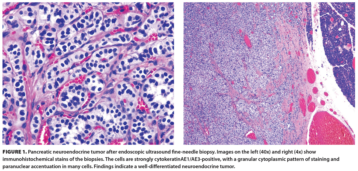
Pancreatic NETs can be divided into well-differentiated NETs (grades 1 to 3),[13] neuroendocrine carcinomas (small cell or large cell), and mixed neuroendocrine–nonneuroendocrine neoplasms [Table]. Mixed lesions have both neuroendocrine and nonneuroendocrine components.[11] The most common nonneuroendocrine component is adenocarcinoma.[11] Histologically, well-differentiated NETs are monomorphic neoplastic proliferations with salt-and-pepper chromatin most often arranged in trabeculae and nests [Figure 1]. These lesions also have a Ki-67 proliferation rate of less than 50%.[11,15,16] Common molecular alterations in well-differentiated NETs are in the MEN1, DAXX, ATRX, mTOR, CDKN2A, and SET2 genes.[17] All pancreatic NETs are considered malignant; however, small, well-differentiated T1 tumors have an excellent prognosis.[11]
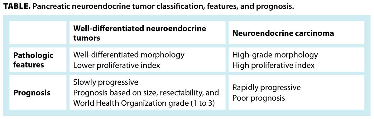
Neuroendocrine carcinomas are distinct malignancies from the well-differentiated NETs [Table]. They can be divided into small-cell and large-cell carcinomas. Small-cell carcinomas are characterized by closely packed cells with a high nuclear-to-cytoplasmic ratio, hyperchromasia, nuclear molding, high mitotic rate, and abundant apoptosis. Large-cell neuroendocrine carcinomas show large, closely packed cells with vesicular chromatin and a prominent nucleolus. The threshold between high-grade (grade 3) well-differentiated NETs and large-cell neuroendocrine carcinomas can be difficult. Rarely, some well-differentiated NETs transform into neuroendocrine carcinomas.[16-18] Neuroendocrine carcinomas frequently show mutations in both the p53 and Rb1 genes. They may also demonstrate gene mutations seen in pancreatic adenocarcinoma, such as KRAS, CDKN2A, and SMAD4 mutations.[17]
Diagnostic evaluation
The clinical presentation of pancreatic NETs can be diverse. Insulinomas can cause intermittent hypoglycemia. Abdominal pain, heartburn, peptic ulcers, or diarrhea can direct clinicians to look for a gastrinoma in Zollinger-Ellison syndrome. VIPomas can cause a rare syndrome of hypokalemia, hypochlorhydria, and watery diarrhea. Nonfunctioning pancreatic NETs can cause complications such as weight loss, abdominal pain, and jaundice from mass effect. Nonfunctioning pancreatic NETs are often found when imaging is being done for another indication.
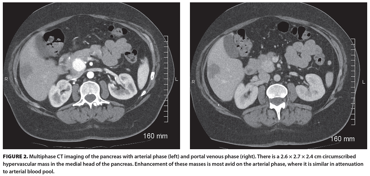
A thorough elevation of pancreatic NETs starts with imaging, includes a functional assessment, and is confirmed by cytology or histology. Routine CT with portal venous phase may miss a pancreatic NET, because the lesion can appear isodense or only mildly hyperdense to the background parenchyma.[19] On arterial phase CT, these lesions demonstrate arterial phase hyperenhancement.[14] Therefore, multiphase cross-sectional imaging is essential to define these lesions [Figure 2]. On MRI, the lesions are hypointense on intrinsic T1 phases and high signal on T2-weighted sequences and demonstrate avid arterial phase enhancement on postcontrast T1 sequences.[20,21] Other arterial enhancing lesions can mimic pancreatic NETs on CT, including solid pseudopapillary neoplasms, pancreatic metastases (e.g., renal cell carcinoma), acinar cell carcinomas, pancreatic hamartomas, pseudoaneurysms, intrapancreatic splenules, and exophytic gastrointestinal stromal tumors.[22,23] The most appropriate way to initiate investigation of a pancreatic lesion is a pancreatic-protocol CT scan.
Depending on the diagnostic evaluation, further noninvasive imaging may be indicated, including an octreotide scan or PET. Three PET radiopharmaceuticals are available for suspected pancreatic NETs: gallium-68 DOTA-Tyr(3)-octreotate (Ga-68 DOTATATE), gallium-68 DOTA-D-Phe1-Tyr(3)-octreotide (Ga-68 DOTATOC), and copper-64 DOTA-Tyr(3)-octreotate (Cu-64 DOTATATE).[24]
Functional testing with biomarkers is usually performed by a local specialist in endocrinology, gastroenterology, hepatopancreaticobiliary surgery, or oncology. The specialist interrogates symptoms, elicits a family history, and examines the patient for signs of multiple endocrine neoplasia type 1, von Hippel-Lindau disease, neurofibromatosis type 1, or tuberous sclerosis complex.[13] North American guidelines recommend against routine use of nonspecific tumor markers such as chromogranin A.[13]
Endoscopic ultrasound
In British Columbia, endoscopic ultrasound and fine-needle biopsy are available at Victoria General Hospital, Vancouver General Hospital, St. Paul’s Hospital, Royal Columbian Hospital, Surrey Memorial Hospital, Royal Inland Hospital, and Kelowna General Hospital. Endoscopic ultrasound has good accuracy for pancreatic NETs[26,27] and facilitates tissue acquisition for most patients.[28] Endoscopic ultrasound is more sensitive than CT[29] and is superior to MRI and somatostatin receptor scintigraphy for pancreatic NET localization; it has reported sensitivities of 82% to 93% and is recommended in guidelines for pancreatic NET diagnosis.[30] Even small pancreatic NETs should be confirmed on biopsy if possible.[31] Endoscopic ultrasound prior to surgery is beneficial. A meta-analysis of 612 patients showed a preoperative sensitivity for pancreatic NET detection of 97%.[32]
For small nonfunctioning pancreatic NETs with low proliferative activity, surveillance with endoscopic ultrasound or cross-sectional imaging is often appropriate. Novel technologies in endoscopic ultrasound that use IV contrast, tissue harmonics, and elastography may be useful in differentiating pancreatic NETs from benign pancreatic lesions in the future. Endoscopic ultrasound can also play a role in the treatment of pancreatic NETs using endoscopic ultrasound–guided tattooing or fiducial insertion to target radiotherapy and surgical treatment. For nonsurgical candidates, endoscopic ultrasound–guided ablation therapy with ethanol and radiofrequency ablation have been studied for local tumor control.[33]
Prognosis
Pancreatic NETs encompass a spectrum of disease, ranging from low-grade indolent tumors to rapidly progressive disease. Tumor histology, grade, Ki-67 score, and clinical stage are important features for estimating prognosis and response to treatment. For localized, resectable disease, 5-year overall survival rates are 97%, 87%, and 73% for stages I, II, and III, respectively, by European Society of Neuroendocrine Tumors staging.[34] Ki-67 is a protein found in dividing cells and is useful for quantifying the risk of tumor growth and spread. Tumor grade, Ki-67 score, and differentiation are strong predictors of prognosis.[35] Based on surveillance, epidemiology, and end results data for patients with stage IV or metastatic disease, 5-year overall survival rates are 51%, 45%, and 12% for tumor grades 1, 2, and 3, respectively.[6] In contrast, pancreatic neuroendocrine carcinomas have a poor prognosis, with a median overall survival of 8 to 17 months.[36] Therefore, when counseling a patient regarding a diagnosis of pancreatic NET, it is important to incorporate both clinical and pathologic features.
Management
Surgery is advised for functional pancreatic NETs.[25] Active surveillance with annual imaging has been advised for patients with nonfunctional pancreatic NETs that are less than 1 cm in size.[25,31] In a prospective study of patients with documented pancreatic NETs that were less than or equal to 2 cm, only 11% of patients showed an increase in pancreatic NET size of at least 0.5 cm/year, which was a study indication for surgical referral, over a median follow-up of 17 months.[37] The management of patients with nonfunctional pancreatic NETs 1 cm to 2 cm, without pancreatic duct obstruction, can be tailored based on surgical risks and tumor biology. Referrals for surgery should be made when the nonfunctional pancreatic NET is 2 cm or larger or is associated with main pancreatic duct dilation.[25] Curative intent surgery for nonfunctional pancreatic NET has a 5-year survival rate of more than 70%.[38] Given the potential for late recurrences, the North American Neuroendocrine Tumor Society recommends surveillance imaging for at least 10 years after resection. Individual recommendations are based on tumor size, Ki-67 score, and the number of lymph nodes involved.[13]
Systemic therapy
Systemic therapy is reserved for patients with unresectable or metastatic disease. Currently, there is no evidence to support the use of systemic therapy in the adjuvant setting. The choice of systemic therapy depends on symptoms, somatostatin receptor status, tumor grade, and histology.
For patients with well-differentiated tumors, a Ki-67 score of less than 10%, and somatostatin receptor–positive disease, somatostatin analogues alone can be considered for first-line therapy with long-term responses reported.[39] However, for patients with well-differentiated pancreatic NETs and a high proliferation rate (Ki-67 > 10%), chemotherapy with capecitabine and temozolomide can be considered.[40,41] New data support the use of peptide receptor radionuclide therapy as first-line treatment in patients with a Ki-67 score of 20% or higher in combination with a somatostatin analogue or as second-line therapy for patients whose disease has progressed on somatostatin analogues.[42,43] Subsequent lines of therapy include tyrosine kinase inhibitors such as sunitinib, cabozantinib, or a mechanistic target of rapamycin inhibitors such as everolimus.[44-46]
In contrast, pancreatic neuroendocrine carcinomas are associated with aggressive behavior and poor prognosis. Neuroendocrine carcinomas are treated with cisplatin and etoposide or fluoropyrimidine-based doublet chemotherapy.
Summary
Pancreatic malignancy is a challenging topic to discuss with patients. This is particularly true if there is a delay in diagnosis. Most patients do not interact with an active group of advocates and survivors. Advocacy programs for other cancers have improved outcomes.[47] Finding the balance between informing a patient and not creating panic is a constant focus for physicians in this field.
Accurate prognostication and awareness of the stark contrast between the management of pancreatic NETs and that of pancreatic adenocarcinoma are critical. A pancreatic-protocol CT with arterial phase imaging is the first step in evaluating a patient with a suspected pancreatic NET. Subsequent referral for endoscopic ultrasound will enable tissue diagnosis and risk stratification. Endoscopic ultrasound providers will refer the patient to a pancreas surgeon or to BC Cancer based on staging data and functional testing. For oncologists and surgeons, deciding between active surveillance with yearly imaging and surgical resection often requires a multidisciplinary review.
Key points
- A pancreatic-protocol CT is the starting imaging modality for a suspected pancreatic neuro-endocrine tumor (NET).
- Functional testing should be initiated by local specialists in endocrinology, gastroenterology, hepatopancreaticobiliary surgery, and oncology.
- A referral for a local endoscopic ultrasound can facilitate detection, tissue diagnosis, and risk stratification.
- The decision between surveillance and surgery is dictated by clinical factors, pancreatic NET size, functional status, stage, and patient goals.
Competing interests
Dr De Korompay has been a speaker and consultant for Penumbra, Inc. and Cook Medical. The remaining authors have no competing interests to declare.
This article has been peer reviewed.

This work is licensed under a Creative Commons Attribution-NonCommercial-NoDerivatives 4.0 International License.
References
1. Dasari A, Shen C, Halperin D, et al. Trends in the incidence, prevalence, and survival outcomes in patients with neuroendocrine tumors in the United States. JAMA Oncol 2017;3:1335-1342. https://doi.org/10.1001/jamaoncol.2017.0589.
2. Hallet J, Law CHL, Cukier M, et al. Exploring the rising incidence of neuroendocrine tumors: A population-based analysis of epidemiology, metastatic presentation, and outcomes. Cancer -2015;121:589-597. https://doi.org/10.1002/cncr.29099.
3. Moldow RE, Connelly RR. Epidemiology of pancreatic cancer in Connecticut. Gastroenterology 1968;55:677-686.
4. Yao JC, Eisner MP, Leary C, et al. Population-based study of islet cell carcinoma. Ann Surg Oncol 2007;14:3492-3500. https://doi.org/10.1245/s10434-007-9566-6.
5. Vagefi PA, Razo O, Deshpande V, et al. Evolving patterns in the detection and outcomes of pancreatic neuroendocrine neoplasms: The Massachusetts General Hospital experience from 1977 to 2005. Arch Surg 2007;142:347-354. https://doi.org/10.1001/archsurg.142.4.347.
6. Sonbol MB, Mazza GL, Mi L, et al. Survival and incidence patterns of pancreatic neuroendocrine tumors over the last 2 decades: A SEER database analysis. Oncologist 2022;27:573-578. https://doi.org/10.1093/oncolo/oyac049.
7. Bruzoni M, Johnston E, Sasson AR. Pancreatic incidentalomas: Clinical and pathologic spectrum. Am J Surg 2008;195:329-332. https://doi.org/10.1016/j.amjsurg.2007.12.027.
8. Heitz PU, Komminoth P, Perren A, et al. Pancreatic endocrine tumors: Introduction. In: WHO classification of tumours. 3rd ed. Vol 8: Pathology and genetics of tumors of endocrine organs. DeLellis RA, Lloyd RV, Heitz PU, Eng C, editors. Lyon, France: International Agency for Research on Cancer; 2004. pp. 177-182.
9. Geurts JL. Inherited syndromes involving pancreatic neuroendocrine tumors. J Gastrointest Oncol 2020;11:559-566. https://doi.org/10.21037/jgo.2020.03.09.
10. Ehehalt F, Saeger HD, Schmidt CM, Grützmann R. Neuroendocrine tumors of the pancreas. Oncologist 2009;14:456-467. https://doi.org/10.1634/theoncologist.2008-0259.
11. Taskin OC, Clarke CN, Erkan M, et al. Pancreatic neuroendocrine neoplasms: Current state and ongoing controversies on terminology, classification, and prognostication. J Gastrointest Oncol 2020;11:548-558.
12. Hofland J, Falconi M, Christ E, et al. European Neuroendocrine Tumor Society 2023 guidance paper for functioning pancreatic neuroendocrine tumour syndromes. J Neuroendocrinol 2023;35:e13318. https://doi.org/10.1111/jne.13318.
13. Halfdanarson TR, Strosberg JR, Tang L, et al. The North American Neuroendocrine Tumor Society consensus guidelines for surveillance and medical management of pancreatic neuroendocrine tumors. Pancreas 2020;49:863-881. https://doi.org/10.1097/MPA.0000000000001597.
14. Ciaravino V, De Robertis R, Tinazzi Martini P, et al. Imaging presentation of pancreatic neuroendocrine neoplasms. Insights Imaging 2018;9:943-953. https://doi.org/10.1007/s13244-018-0658-6.
15. Klimstra DS, Klöppel G, La Rosa S, et al. Classification of neuroendocrine neoplasms of the digestive system. In: WHO classification of tumours. 5th ed. Vol 1: Digestive system tumours. WHO Classification of Tumours Editorial Board, editor. Lyon, France: International Agency for Research on Cancer; 2019. pp. 16-19.
16. Tang LH, Basturk O, Sue JJ, Klimstra DS. A practical approach to the classification of WHO grade 3 (G3) well-differentiated neuroendocrine tumor (WD-NET) and poorly differentiated neuroendocrine carcinoma (PD-NEC) of the pancreas. Am J Surg Pathol 2016;40:1192-1202. https://doi.org/10.1097/PAS.0000000000000662.
17. Umetsu SE, Kakar S, Basturk O, et al. Integrated genomic and clinicopathologic approach distinguishes pancreatic grade 3 neuroendocrine tumor from neuroendocrine carcinoma and identifies a subset with molecular overlap. Mod Pathol 2023;36:100065.
18. Basturk O, Yang Z, Tang LH, et al. The high-grade (WHO G3) pancreatic neuroendocrine tumor category is morphologically and biologically heterogenous and includes both well differentiated and poorly differentiated neoplasms. Am J Surg Pathol 2015;39:683-690. https://doi.org/10.1097/PAS.0000000000000408.
19. Kartalis N, Mucelli RMP, Sundin A. Recent developments in imaging of pancreatic neuroendocrine tumors. Ann Gastroenterol 2015;28:193-202.
20. Lo GC, Kambadakone A. MR imaging of pancreatic neuroendocrine tumors. Magn Reson Imaging Clin N Am 2018;26:391-403. https://doi.org/10.1016/j.mric.2018.03.010.
21. Sahani DV, Bonaffini PA, Fernández-Del Castillo C, Blake MA. Gastroenteropancreatic neuroendocrine tumors: Role of imaging in diagnosis and management. Radiology 2013;266:38-61. https://doi.org/10.1148/radiol.12112512.
22. Raman SP, Hruban RH, Cameron JL, et al. Pancreatic imaging mimics: Part 2, pancreatic neuroendocrine tumors and their mimics. Am J Roentgenol 2012;199:309-318. https://doi.org/10.2214/AJR.12.8627.
23. Bhosale PR, Menias CO, Balachandran A, et al. Vascular pancreatic lesions: Spectrum of imaging findings of malignant masses and mimics with pathologic correlation. Abdom Imaging 2013;38:802-817. https://doi.org/10.1007/s00261-012-9954-7.
24. Johnbeck CB, Knigge U, Loft A, et al. Head-to-head comparison of 64Cu-DOTATATE and 68Ga-DOTATOC PET/CT: A prospective study of 59 patients with neuroendocrine tumors. J Nucl Med 2017;58:451-457. https://doi.org/10.2967/jnumed.116.180430.
25. Howe JR, Merchant NB, Conrad C, et al. The North American Neuroendocrine Tumor Society consensus paper on the surgical management of pancreatic neuroendocrine tumors. Pancreas 2020;49:1-33. https://doi.org/10.1097/MPA.0000000000001454.
26. Puli SR, Kalva N, Bechtold ML, et al. Diagnostic accuracy of endoscopic ultrasound in pancreatic neuroendocrine tumors: A systematic review and meta analysis. World J Gastroenterol 2013;19:3678-3684. https://doi.org/10.3748/wjg.v19.i23.3678.
27. Iabichino G, Di Leo M, Arena M, et al. Diagnosis, treatment, and current concepts in the endoscopic management of gastroenteropancreatic neuroendocrine neoplasms. World J Gastroenterol 2022;28:4943-4958. https://doi.org/10.3748/wjg.v28.i34.4943.
28. Pais SA, Al-Haddad M, Mohamadnejad M, et al. EUS for pancreatic neuroendocrine tumors: A single-center, 11-year experience. Gastrointest Endosc 2010;71:1185-1193. https://doi.org/10.1016/j.gie.2009.12.006.
29. Khashab MA, Yong E, Lennon AM, et al. EUS is still superior to multidetector computerized tomography for detection of pancreatic neuro-endocrine tumors. Gastrointest Endosc 2011;73:691-606. https://doi.org/10.1016/j.gie.2010.08.030.
30. Eloubeidi MA, Decker GA, Chandrasekhara V, et al. The role of endoscopy in the evaluation and management of patients with solid pancreatic neoplasia. Gastrointest Endosc 2016;83:17-28.
31. Kos-Kudła B, Castaño JP, Denecke T, et al. European Neuroendocrine Tumour Society (ENETS) 2023 guidance paper for nonfunctioning pancreatic neuroendocrine tumours. J Neuroendocrinol 2023;35:e13343. https://doi.org/10.1111/jne.13343.
32. James PD, Tsolakis AV, Zhang M, et al. Incremental benefit of preoperative EUS for the detection of pancreatic neuroendocrine tumors: A meta-analysis. Gastrointest Endosc 2015;81:848-856.e1. https://doi.org/10.1016/j.gie.2014.12.031.
33. Melita G, Pallio S, Tortora A, et al. Diagnostic and interventional role of endoscopic ultrasonography for the management of pancreatic neuroendocrine neoplasms. J Clin Med 2021;10:2638. https://doi.org/10.3390/jcm10122638.
34. Ellison TA, Wolfgang CL, Shi C, et al. A single institution’s 26-year experience with nonfunctional pancreatic neuroendocrine tumors: A validation of current staging systems and a new prognostic nomogram. Ann Surg 2014;259:204-212. https://doi.org/10.1097/SLA.0b013e31828f3174.
35. Yang M, Ke N-w, Zhang Y, et al. Survival analyses for patients with surgically resected pancreatic neuroendocrine tumors by World Health Organization 2010 grading classifications and American Joint Committee on Cancer 2010 staging systems. Medicine (Baltimore) 2015;94:e2156. https://doi.org/10.1097/MD.0000000000002156. Erratum in: Medicine (Baltimore) 2016;95:e507d. https://doi.org/10.1097/01.md.0000480962.17450.7d.
36. Kaslow SR, Vitiello GA, Prendergast K, et al. Surgical treatment of patients with poorly differentiated pancreatic neuroendocrine carcinoma: An NCDB analysis. Ann Surg Oncol 2022;29:3522-3531. https://doi.org/10.1245/s10434-022-11477-5.
37. Heidsma CM, Engelsman AF, van Dieren S, et al. Watchful waiting for small non-functional pancreatic neuroendocrine tumours: Nationwide prospective cohort study (PANDORA). Br J Surg 2021;108:888-891. https://doi.org/10.1093/bjs/znab088.
38. Ricci C, Partelli S, Landoni L, et al. Sporadic non-functioning pancreatic neuroendocrine tumours: Multicentre analysis. Br J Surg 2021;108:811-816. https://doi.org/10.1093/bjs/znaa141.
39. Caplin ME, Pavel M, Ruszniewski P. Lanreotide in metastatic enteropancreatic neuroendocrine tumors. N Engl J Med 2014;371:1556-1557. https://doi.org/10.1056/NEJMc1409757.
40. Strosberg JR, Fine RL, Choi J, et al. First-line chemotherapy with capecitabine and temozolomide in patients with metastatic pancreatic endocrine carcinomas. Cancer 2011;117:268-275. https://doi.org/10.1002/cncr.25425.
41. Kunz PL, Graham NT, Catalano PJ, et al. Randomized study of temozolomide or temozolomide and capecitabine in patients with advanced pancreatic neuroendocrine tumors (ECOG-ACRIN E2211). J Clin Oncol 2023;41:1359-1369. https://doi.org/10.1200/JCO.22.01013. Epub 2022 Oct 19.
42. Singh S, Halperin D, Myrehaug S, et al. [177Lu]Lu-DOTA-TATE plus long-acting octreotide versus high‑dose long-acting octreotide for the treatment of newly diagnosed, advanced grade 2–3, well-differentiated, gastroenteropancreatic neuroendocrine tumours (NETTER-2): An open-label, randomised, phase 3 study. Lancet 2024;403(10446):2807-2817. https://doi.org/10.1016/S0140-6736(24)00701-3.
43. Baudin E, Walter TA, Beron A, et al. First multicentric randomized phase II trial investigating the antitumor efficacy of peptide receptor radionucleide therapy with 177Lutetium-Octreotate (OCLU) in unresectable progressive neuroendocrine pancreatic tumor: Results of the OCLURANDOM trial. Ann Oncol 33:S95. https://doi.org/10.1016/j.annonc.2022.07.1013.
44. Yao JC, Shah MH, Ito T, et al. Everolimus for advanced pancreatic neuroendocrine tumors. N Engl J Med 2011;364:514-523. https://doi.org/10.1056/NEJMoa1009290.
45. Raymond E, Dahan L, Raoul J-L, et al. Sunitinib malate for the treatment of pancreatic neuroendocrine tumors. N Engl J Med 2011;364:501-513. https://doi.org/10.1056/NEJMoa1003825. Erratum in N Engl J Med 2011;364:1082.
46. Chan JA, Geyer S, Zemla T, et al. Phase 3 trial of cabozantinib to treat advanced neuroendocrine tumors. N Engl J Med 2025;392:653-665. https://doi.org/10.1056/NEJMoa2403991.
47. Schliemann D, Htay MNN, Dahlui M, et al. Impact of a mass media campaign on breast cancer symptoms awareness and screening uptake in Malaysia: Findings from a quasi-experimental study. BMJ Open 2020;10:e036503. https://doi.org/10.1136/bmjopen-2019-036503.
Dr Sanders is a gastroenterologist at Kelowna General Hospital and a clinical instructor in the Faculty of Medicine at the University of British Columbia. Dr Berg is a pathologist at Kelowna General Hospital and a clinical instructor in the Faculty of Medicine at UBC. Dr De Korompay is a radiologist at Kelowna General Hospital and a clinical instructor in the Faculty of Medicine at UBC. Dr Nguyen is a medical oncologist at BC Cancer – Kelowna (Sindi Ahluwalia Hawkins Centre) and a clinical assistant professor in the Faculty of Medicine at UBC. Dr Trasolini is a gastroenterologist at Vancouver General Hospital and a clinical assistant professor in the Faculty of Medicine at the UBC.