Clinical physiology of chronic obstructive pulmonary disease
Issue: BCMJ,
vol. 50 , No. 2 , March 2008 ,
Pages 97-102 Clinical Articles
The symptoms and functional limitations of chronic obstructive pulmonary disease (COPD) are a direct result of airway and lung parenchymal processes. These processes lead to airflow obstruction, increased work of breathing, and gas exchange abnormalities. When advanced, these can in turn cause pulmonary hypertension, cor pulmonale, and left heart dysfunction. Cataloguing of symptoms, physical examination, and routine radiology are of limited utility in the early diagnosis and assessment of response to therapy for COPD. Patients can benefit from the use of laboratory measurements to manage this disorder. Spirometry and oximetry have the broadest application, while arterial blood gas and exercise testing have selected utility.
A variety of methods can be used to assess disturbances and dysfunctions common in COPD, including pulse oximetry and exercise testing.
Although many different factors may be involved in the development of COPD, smoking is the most significant. Exposure to tobacco smoke provokes a series of inflammatory processes involving the small as well as the large airways and can also impair host defence mechanisms.
In predisposed individuals this leads to pathological processes that we have come to recognize as chronic bronchitis and emphysema. It is important to note that bronchial inflammation can be detected long before the disease is evident clinically[1] and persists long after the cessation of smoking,[2] and that the inflammatory processes of COPD are readily distinguishable from those of asthma.[3,4]
Physiological defects
Airflow limitation and hyperinflation
Expiratory airflow limitation is the principal physiological defect in COPD. Intrinsic airway factors relate to bronchial wall inflammation and include mucosal inflammation/edema, bronchial wall remodeling/fibrosis, and increased mucosal secretions.
Extrinsic factors involve the loss of elastic tissue support for small airways and the dynamic expiratory compression of these airways.[5,6] Other factors such as respiratory muscle dysfunction can further limit airflow in some patients.
Hyperinflation can also occur in COPD, leading to an increase in functional residual capacity (FRC)—the amount of air that remains in the lungs at the end of tidal exhalation.
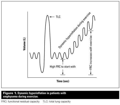
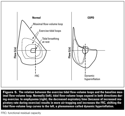
This can lead to gas trapping and an increase in residual volume (RV). As a consequence, there is an augmentation of the inspiratory work of breathing, which is an important factor in producing dyspnea. As the expiratory time is essential for lung emptying, factors that decrease this time, such as an increasing respiratory rate during exercise, result in a progressive increase in FRC (Figure 1).[7] This phenomenon is called dynamic hyperinflation, and is largely responsible for exercise limitation in COPD.[7-11]
Gas exchange disturbances
Perturbations in gas exchange are caused primarily by regional inequalities of ventilation and perfusion (VQ mismatching).[12] This process commonly produces hypoxemia, but in more advanced disease can also contribute to hypercapnia and chronic respiratory acidosis. Other contributing factors to gas exchange disturbances in advanced COPD are pulmonary hypertension and impaired cardiac function, which can lead to reduced mixed venous oxygenation.
Ventilatory muscle dysfunction
A number of factors contribute to ventilatory muscle dysfunction in COPD. A major factor is a consequence of hyperinflation, which limits force generation and endurance,[13] and places the inspiratory muscles at a mechanical disadvantage.
Other factors include nutritional alterations, a sustained inflammatory response that affects the contractile apparatus, tissue hypoxia, and loss of muscle mass.[14,15] These factors also affect other skeletal muscles, which may further contribute to exercise limitation.[16,17]
Cardiovascular disturbances
Cardiovascular disturbances are common in COPD and may represent a complication of COPD itself or may be triggered by the same factor, that is, smoking. Recently it has been proposed that lung inflammation may directly affect atherogenesis by driving systemic inflammation.
Pulmonary hypertension is a late complication of COPD and independently worsens its prognosis.[18-24] A major factor is chronic hypoxia, which can result in pulmonary vasoconstriction. Other factors include endothelial dysfunction, remodeling of pulmonary arteries, and pulmonary capillary bed destruction.
Right ventricular dysfunction and failure (cor pulmonale) may eventually develop and add to the morbidity and mortality of this disease.
Reduced exercise capacity
Deconditioning may result from lack of physical activity and can be an independent factor in exercise limitation. Fortunately, this is amenable to a pulmonary rehabilitation program. Management of deconditioning, like the management of other physiological defects of COPD, begins with assessment of function.
Assessment of airflow limitation and hyperinflation
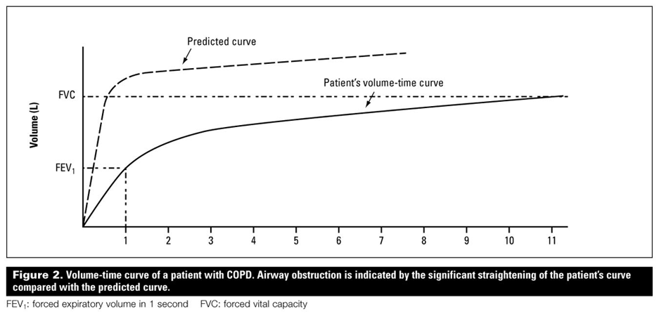
Spirometry with measurements derived from a maximal forced expiratory maneuver is the most clinically relevant test in assessing COPD. Airway obstruction is diagnosed when the FEV1/FVC (forced expiratory volume in 1 second to forced vital capacity) ratio is less than 0.7 L, a situation usually accompanied by a reduction in FEV1 to less than 80% of predicted (Figure 2).
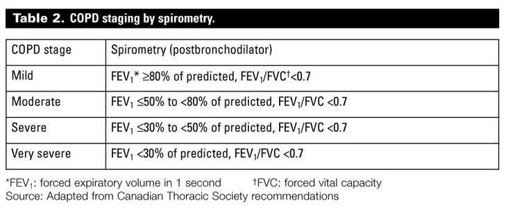
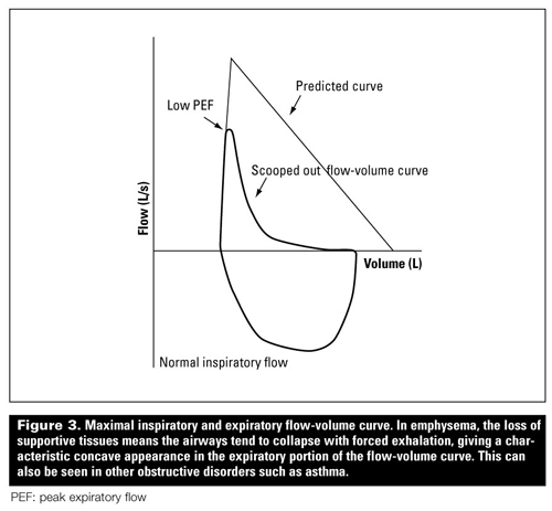
FEV1 has traditionally been used to grade severity of COPD (see Table 2 from Pharmacological management of stable chronic obstructive pulmonary disease).[25-26] In COPD the expiratory flow-volume curve is characteristically concave (Figure 3). This appearance is typical for emphysema (but not specific) and implies the dynamic airway collapse with forced exhalation.[27] Significant response to bronchodilators, defined as an increase in FEV1 by 12% and 180 mL, is common in asthma but uncommon in COPD.
New technology has allowed the development of portable hand-held spirometers that are robust, simple to use, and reasonable in cost ($500 to $1500), making spirometry a test that can be performed in the office setting. Accreditation can be obtained in BC through the Diagnostic Accreditation Program, and once you have the required pulmonary function test (PFT) credentials you may bill for performing this test. Spirometry should be routine in COPD for the following:
• Diagnosis (particularly of early disease or to differentiate from asthma).
• Establishing severity.
• Determining prognosis.
• Monitoring disease progression and response to therapy.
More detailed lung function tests are available in most PFT labs. These are utilized in select instances in COPD. Most commonly this occurs when there is a reduction in FVC and a need to determine whether there is a concomitant restrictive disorder such as interstitial lung disease. In pure COPD, FVC is modestly decreased if there is air trapping and hyperinflation (RV increased).
Diffusing capacity (DLco) is very sensitive in detecting gas exchange abnormalities. It is usually reduced in emphysema (whereas it is normal or high in asthma) and correlates reasonably well with the pathological severity of emphysema.[28]
Full pulmonary function tests should therefore be considered when spirometry indicates a reduced FVC to determine if there is a restrictive disorder present, and when spirometry is normal despite the presence of dyspnea to determine if there is an impairment in DLco that might suggest another pulmonary parenchymal or vascular disorder.
Peak expiratory flow rate
Peak expiratory flow rate (PEFR) testing provides a simple, readily available measure of airflow. PEFR, however, may underestimate airflow obstruction in COPD as it is predominantly a measure for the large airways. Furthermore, the reproducibility and reliability of PEFR testing is inferior to spirometry. We therefore advise against its use for routine assessment of COPD (in contrast to asthma) although it may have some merit as a serial measurement for a given individual.[29-30]
Radiological tests
Chest radiography may show features of hyperinflation such as flattening of the diaphragms with increased anteroposterior diameter on a lateral film, but this is not sensitive or specific for COPD. Although not routinely indicated, a CT chest scan may on occasion be of use in the characterization of COPD (emphysema).[31]
Assessment of gas exchange disturbances
Pulse oximetry can be done in the ambulatory setting to reliably quantitate oxygen saturation. Arterial blood gas measurement can be considered in patients with Sao2 of less than 92% (or FEV1 of less than 40%) to more precisely assess the degree of hypoxemia and hypercapnia.[32]
Both tests can be used to determine the potential role of domiciliary oxygen. The portability of pulse oximetry makes it applicable in the quantitation of hypoxemia with exertion and during sleep.
Assessment of ventilatory muscle dysfunction
Tests for ventilatory muscle function may be considered in select cases, including those where ventilatory muscle involvement is suspected in a neuromuscular disorder, or where dyspnea or hypercapnia are out of proportion to the patient’s FEV1.[12] Other assessment methods for muscle dysfunction may include measuring lung volumes (restrictive disorder), blood gases (elevated Paco2), and maximal inspiratory and expiratory pressures. EMG and nerve conduction studies may also be considered.
Assessment of cardiovascular disturbances
Clinical evaluation, chest X-ray, EKG, and echocardiography with Echo-Doppler estimation can be used in patients with suspected cardiac disorders or pulmonary hypertension to assess the pulmonary artery systolic pressure and ventricular function.[27]
Assessment of reduced exercise capacity
Dyspnea scale
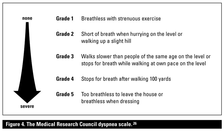
The Medical Research Council dyspnea scale is a simple clinical tool used to assess shortness of breath and disability during activity (Figure 4).[25] This scale has been found to correlate with the patient’s quality of life and may be of use in predicting mortality.[25]
6-minute walk test
The 6-minute walk test is a standardized lab test in which the patient walks unassisted for 6 minutes and the distance is measured. The normal distances walked are about 500 m for women and about 580 m for men, with a change of 40 m in a properly performed test considered significant. The 6-minute walk test distance (degree of reduction) correlates well with functional disability and provides prognostic information.[33-34]
Cardiopulmonary exercise testing
The cardiopulmonary exercise test (CPET) objectively measures maximum exercise capacity. When exercise capacity is reduced, CPET can assist in identifying the organ system responsible. The measure of peak oxygen uptake during incremental testing is an independent prognostic factor in COPD.[20]
Dynamic hyperinflation can be demonstrated by performing repeated inspiratory capacity maneuvers and by comparing the exercise tidal flow-volume loops with the baseline maximal flow-volume loop (Figure 5). CPET can assist with patient selection for pulmonary rehabilitation, lung volume reduction surgery, and suitability for pulmonary resection.
Conclusions
COPD is an inflammatory airway disease with a complex pathophysiology. This disorder leads to airway obstruction and often to hyperinflation, which along with gas exchange abnormalities interact to cause dyspnea and functional limitation. Spirometry is a widely available tool that quantifies the abnormalities in COPD and can be fundamental in diagnosing, monitoring, and establishing the prognosis of individuals with this condition.
More detailed lung function testing may have utility, but is not generally required. Measures of oxygenation (oximetry and arterial blood gases) can be used to monitor disease severity and to determine the appropriateness of domicillary oxygen. Such measurements should play as fundamental a role in the management of COPD as the monitoring of blood sugar does in diabetes.
Competing interests
None declared.
References
1. Hogg JC, Chu F, Utokaparch S, et al. The nature of small-airway obstruction in chronic obstructive pulmonary disease. N Engl J Med 2004;350:2645-2653.
2. Shapiro SD. End-stage chronic obstructive pulmonary disease. The cigarette is burned out but inflammation rages on. Am J Respir Crit Care Med 2001;164:339-340.
3. Saetta M, Di Stefano A, Turato G, et al. CD8+ T-lymphocytes in peripheral airways of smokers with chronic obstructive pulmonary disease. Am J Respir Crit Care Med 1998;157:822-826.
4. Fabbri LM, Romagnoli M, Corbetta L, et al. Differences in airway inflammation in patients with fixed airflow obstruction due to asthma or chronic obstructive pulmonary disease. Am J Respir Crit Care Med 2003;167:418-424.
5. Leopold JG, Geoff J. Centrilobular form of hypertrophic emphysema and its relation to chronic bronchitis. Thorax 1957;12:219-235.
6. McLean KA. Pathogenesis of pulmonary emphysema. Am J Med 1958;25:62-74.
7. O’Donnell DE, Webb KA. Exertional breathlessness in patients with chronic airflow limitation. The role of lung hyperinflation. Am Rev Respir Dis 1993;48:1351-1357.
8. O’Donnell DE, Revill S, Webb KA. Dynamic hyperinflation and exercise intolerance in chronic obstructive pulmonary disease. Am J Respir Crit Care Med 2001;164:770-777.
9. Tantucci C, Duguet A, Similowski T, et al. Effect of salbutamol on dynamic hyperinflation in chronic obstructive pulmonary disease patients. Eur Respir J 1998;12:799-804.
10. Parker C, Voduc N, Aaron SD, et al. Physiological changes during symptom recovery from moderate exacerbations of COPD. Eur Respir J 2005;26:420-428.
11. Stevenson NJ, Walker PP, Costello RW, et al. Lung mechanics and dyspnea during exacerbations of chronic obstructive pulmonary disease. Am J Respir Crit Care Med 2005;172:1510-1516.
12. Rodriguez-Roisin R, MacNee W. Pathophysiology of chronic obstructive pulmonary disease. Eur Respir Monograph 1998;3:107-126.
13. Hughes RL, Katz H, Sahgal V, et al. Fiber size and energy metabolites in five separate muscles from patients with chronic obstructive pulmonary disease. Respiration 1983;44:321-328.
14. Satta A, Migliori GB, Spanevello A, et al. Fibre types in skeletal muscles of chronic obstructive pulmonary disease patients related to respiratory function and exercise tolerance. Eur Respir J 1997;10:2853-2860.
15. Wilson DO, Rogers RM, Wright EC, et al. Body weight in chronic obstructive pulmonary disease. The National Institutes of Health Intermittent Positive-Pressure Breathing Trial. Am Rev Respir Dis 1989;139:1435-1438.
16 Schols AM, Slangen J, Volovics L, et al. Weight loss is a reversible factor in the prognosis of chronic obstructive pulmonary disease. Am J Respir Crit Care Med 1998;157:1791-1797.
17. Calverley PMA, Anderson JA, Celli B, et al. for the TORCH investigators. Salmeterol and fluticasone propionate and survival in chronic obstructive pulmonary disease. N Engl J Med 2007;356:775-789.
18. Weitzenblum E, Hirth C, Ducolone A, et al. Prognostic value of pulmonary artery pressure in chronic obstructive pulmonary disease. Thorax 1981;36:752-758.
19. Bishop JM, Cross KW. Physiological variables and mortality in patients with various categories of chronic respiratory disease. Bull Eur Physiopathol Respir 1984;20:495-500.
20. Oga T, Nishimura K, Tsukino M, et al. Analysis of the factors related to mortality in chronic obstructive pulmonary disease: Role of exercise capacity and health status. Am J Respir Crit Care Med 2003;167:544-549.
21. Biernacki W, Flenley DC, Muir AL, et al. Pulmonary hypertension and right ventricular function in patients with COPD. Chest 1988;94:1169-1175.
22. MacNee W. Pathophysiology of cor pulmonale in chronic obstructive pulmonary disease. Part two. Am J Respir Crit Care Med 1994;150:1158-1168.
23. Soriano JB, Visick GT, Muellerova H, et al. Patterns of comorbidities in newly diagnosed COPD and asthma in primary care. Chest 2005; 128:2099-2107.
24. Sidney S, Sorel M, Quesenberry CP Jr, et al. COPD and incident cardiovascular disease hospitalizations and mortality: Kaiser Permanente Medical Care Program. Chest 2005;128:2068-2075.
25. O’Donnell DE, Aaron S, Bourbeau J, et al. Canadian Thoracic Society recommendations for management of chronic obstructive pulmonary disease—2003. Can Respir J 2003;10(suppl A):11A-65A.
26. Hancox R, Whyte K. Pocket Guide to Lung Function Tests. Sydney: McGraw-Hill; 2001. 157 pp.
27. Gibson GJ, MacNee W. Chronic obstructive pulmonary disease: Investigations and assessment of severity. Eur Respir Monograph 2006;11:24-40.
28. McLean A, Warren PM, Gillooly M, et al. Microscopic and macroscopic measurements of emphysema: Relation to carbon monoxide gas transfer. Thorax 1992;47:144-149.
29. BTS guidelines for the management of chronic obstructive pulmonary disease. The COPD Guidelines Group of the Standards of Care Committee of the BTS. Thorax 1997;52(suppl 5):S1-28.
30. Kelly CA, Gibson GJ. Relation between FEV and peak expiratory flow in patients with chronic airflow obstruction. Thorax 1988;43:335-336.
31. Pauwels RA, Buist AS, Ma P, et al. Global strategy for the diagnosis, management, and prevention of chronic obstructive pulmonary disease: National Heart, Lung, and Blood Institute and World Health Organization Global Initiative for Chronic Obstructive Lung Disease (GOLD): Executive summary. Respir Care 2001;46:798-825.
32. Roberts CM, Bugler JR, Melchor R, et al. Value of pulse oximetry in screening for long-term oxygen therapy requirement. Eur Respir J 1993;6:559-562.
33. Pinto-Plata VM, Cote C, Cabral H, et al. The 6-min walk distance: Change over time and value as a predictor of survival in severe COPD. Eur Respir J 2004;23:28-33.
34. Casanova C, Cote CG, Marin JM, et al. The 6-min walking distance: Long-term follow up in patients with COPD. Eur Respir J 2007; 29:535-540.
Dr Al Talag is a fellow in the division of Respirology Medicine at the University of British Columbia.
Dr Wilcox is a respirologist at St. Paul's Hospital, associate professor of medicine at UBC, the director for Respirology Training at UBC, and the medical director of the Pulmonary Function Laboratory St. Paul’s Hospital.
