Cardiac transplantation in British Columbia
Issue: BCMJ,
vol. 52 , No. 4 , May 2010 ,
Pages 197-202 Clinical Articles
The first cardiac transplantation in the world was performed in 1967. Since then it has become standard therapy for end-stage heart failure with over 200 recipients in British Columbia over the past 20 years. Cardiac transplantation should be considered in individuals with advanced heart disease who have a poor chance of long-term survival despite optimal medical or surgical therapy. Contraindications to transplant include pulmonary hypertension, active infection, systemic illness, renal dysfunction, recent malignancy, active smoking/substance abuse, or the inability to undergo rehabilitation. All patients require immunosuppressive therapy following transplantation to prevent allograft rejection. In addition to rejection, common complications in the post-transplant period include infections, malignancy, post-transplant lymphoproliferative disorder, and allograft coronary artery disease. Given the scarcity of available donor hearts in BC, efforts have been made recently to develop mechanical ventricular assist devices to support patients waiting for donor organs.
Standard malignancy screening and aggressive management of atherosclerotic risk factors are both needed after a heart transplant.
In December 1967 Christiaan Barnard performed the first human cardiac transplantation in Cape Town, South Africa. Louis Washkansky, a 55-year-old man, survived for 18 days before succumbing to pneumonia.
Since then cardiac transplantation has evolved to become a widely adopted therapeutic option for the treatment of end-stage heart failure. By 2007 over 80000 heart transplants were reported in the International Society of Heart and Lung Transplant (ISHLT) worldwide data registry.[1]
In BC the first cardiac transplantation was performed in 1988. Since then over 200 patients have undergone transplantation. More than 70% of patients survive the first 5 years following transplant (Figure 1). Increasing recipient age, pre-existing renal dysfunction, and an elevated body mass index adversely affect long-term survival.
At any given time there are 5 to 15 patients on the active transplant list. The average wait list time in British Columbia in 2007 was 141 days (Figure 2), with longer wait times for male patients (average 167 days vs 65 for female patients) and those with a higher body mass index (average time 323 days if BMI>31), or more common blood type (average 223 days if type O).
Having type O blood lengthens the wait list not only because more transplant candidates have this blood type to begin with, but because type O patients can only receive an organ from a type O donor—unlike type AB patients, who are universal recipients.
On average patients spend 2 weeks in the hospital after transplantation, including 3 to 5 days in the ICU. This is followed by weekly outpatient visits and endomyocardial biopsies for the first month. Patients can expect about 12 to 15 biopsies in the first year following surgery. Frequent evaluation is required to ensure graft survival and the prevention of complications. All patients must remain in the Lower Mainland during the first 3 months following transplant.
Indications
Cardiac transplantation should be considered in individuals with advanced heart disease who have a poor chance of long-term survival despite optimal medical or surgical therapy. This includes patients with end-stage heart failure (ischemic or nonischemic), refractory ventricular arrhythmias, or congenital heart defects not amenable to surgical repair.[2]
Predictors of mortality in this patient population include poor left ventricular function, hyponatremia, elevated BNP, ischemic heart disease, elevated resting heart rate, low mean arterial blood pressure, intraventricular conduction defects on ECG, and a low aerobic threshold during cardiopulmonary exercise testing.[3]
Patients with markedly impaired left ventricular function and NYHA class III or IV symptoms should undergo cardiopulmonary exercise testing to objectively assess their functional capacity. Peak oxygen consumption (VO2) less than 10 mL/kg/min is an absolute indication for transplantation. A value between 11 mL/kg/min and 14 mL/kg/min, or less than 55% of the age-predicted peak, is a relative indication.[4]
Individuals who may be considered for cardiac transplantation should be referred to the Pre-Transplant Clinic at St. Paul’s Hospital (Table 1). Patients are required to undergo a series of investigations in order to determine candidacy. These include a chest X-ray, pulmonary function tests, an abdominal ultrasound, a bone density scan, a mammogram (in women over 40), and a carotid duplex scan (if over age 40).
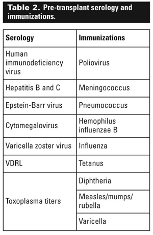
Because of the need for lifelong immunosuppression therapy after transplantation, all candidates must be tested for prior exposure to various pathogens and receive multiple immunizations (Table 2). Typically these investigations are coordinated at the time of the patient’s first visit to the clinic.
Contraindications
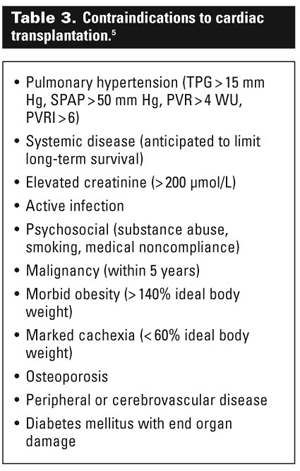
There are multiple contraindications to cardiac transplantation (Table 3).[4] Pulmonary hypertension is one of the most important. Right ventricular dysfunction in the presence of high pulmonary pressures is a common cause of primary graft failure and death following transplant.
All patients require a right heart catheterization during their initial pre-transplant assessment. Pulmonary pressure reversibility should be assessed if the pulmonary artery pressure (PAP) is greater than 50 mm Hg, the transpulmonary gradient is greater than 14 mm Hg (PAP–pulmonary capillary wedge pressure), or the pulmonary vascular resistance (PVR) is more than 3 Wood units.
This usually involves continuous administration of inhaled nitric oxide or infusion of an inotrope such as milrinone, which has vasodilatory properties. The following values represent an absolute contraindication to transplantation:
• PVR>6 Wood units
• Transpulmonary gradient>16 mm Hg
• PVR>2.5 Wood units following a vasodilatory challenge[2]
Additional contraindications include active infection, systemic illness, renal dysfunction (Cr>200), recent malignancy (within 5 years), active smoking/substance abuse, or the inability to undergo rehabilitation following transplantation.
Osteoporosis is a relative contraindication because of the need for steroids as part of the initial immunosuppressive regimen. Age itself is not an absolute contraindication; however, increasing recipient age is associated with decreased survival rates.[1] Furthermore, older patients often have a greater number of comorbidities that may preclude transplant candidacy.
A psychosocial assessment is completed for all potential candidates. The transplant team has a dedicated social worker and psychologist to facilitate this. It is imperative that patients have a strong support network during the initial period following transplantation, during which time multiple outpatient hospital visits are required.
Medical compliance is also imperative to ensure graft survival, meaning that a history of noncompliance is a relative contraindication to candidacy.
Immunosuppressive therapy
All patients require immunosuppressive therapy following transplantation in order to prevent allograft rejection. All donors and recipients are matched for ABO compatibility. HLA matching is not performed; however, a panel-reactive antibody (PRA) screen is performed prior to transplantation.[5]
This test measures the amount of preformed antibodies to a panel of donor lymphocyte HLAs. A PRA greater than 10% is associated with an increased risk of rejection and mortality. Higher PRA titers are common in multiparous females, patients who have received multiple blood transfusions, or those with a ventricular assist device (VAD).[6]
The initial immunosuppression generally consists of prednisone, a calcineurin inhibitor (tacrolimus or cyclosporine), and a purine synthesis inhibitor such as mycophenolate mofetil.[4] In the absence of rejection, prednisone is frequently withdrawn over the first 6 months post-transplant.
Tacrolimus and cyclosporine levels are monitored to ensure therapeutic dosing and to avoid toxic side effects. These may include hypertension, hyperlipidemia, hyperglycemia, tremor, headaches, electrolyte imbalances, hepatotoxicity, gingival hyperplasia, and hypertrichosis.
Many drugs affect the serum concentrations of calcineurin inhibitors. Caution should be taken when prescribing medications such as calcium channel blockers, antifungals, anti-inflammatories, allopurinol, antiseizure medications, and H2-blockers to these patients.[4]
Post-transplant complications
The most common complications post-transplant are rejection, infections, malignancy, post-transplant lymphoproliferative disorder, and allograft coronary artery disease (ACAD).
Rejection accounts for less than 20% of all deaths within the first year following transplant. This value drops to 5% after the third year.[1] The majority of patients with microscopically proven rejection are asymptomatic and only diagnosed at the time of endomyocardial biopsy.
Symptoms consistent but not specific with graft rejection include fever, malaise, reduced exercise tolerance, hypotension, and clinical signs of congestive heart failure. Biopsies are initially performed weekly for the first month and then gradually decrease in frequency until the patient is on a stable, well-established immunosuppressive regimen.
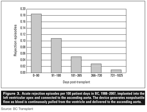
The long-term rates of rejection in British Columbia have decreased over the past 2 decades with the advent of improved regimens. Rejection rates fall steadily after transplant, with the 6 months of the first year posing the highest risk for acute rejection (Figure 3).
The most common opportunistic infections in the first 6 months following transplantation are caused by cytomegalovirus, herpes simplex virus, Pneumocystis carinii (PCP), Aspergillus, Nocardia, and Toxoplasma gondii. All patients receive PCP prophylaxis for the first year following transplant.
Prophylaxis against other opportunistic infections is dependant upon the serologic status of the donor and recipient. Seronegative recipients receive prophylaxis if their donor was seropositive for a given pathogen. After the initial 6 months post-transplant patients remain at risk for common pathogens such as pneumococcal pneumonia and influenza.
An annual flu shot is recommended along with the pneumonia vaccine every 5 years. Shingles is common in this population.
Malignancy is a frequent cause of mortality in patients who survive the first 5 years after transplant. Solid organ or skin cancer occurs in about 13% of patients at 5 years and 30% of patients at 10 years.[7] Skin malignancies are the most common. All patients are advised to use sunscreen and barrier protection while outdoors. An annual skin examination is imperative. Standard age-appropriate screening tests (mammograms, Pap tests, and rectal exams) should be performed.
Post transplant lymphoproliferative disorder refers to a spectrum of diseases from mononucleosis to B-cell monoclonal malignancies. It is associated with Epstein-Barr virus and has many clinical presentations.
Treatment requires reduction or withdrawl of immunosuppressive therapy, and may include the addition of antiviral therapies, intravenous immunoglobulin, or monoclonal antibody therapy such as rituximab.[8] Consultation with hematology or oncology is generally required.
Allograft coronary artery disease is the most common cause of morbidity and mortality following transplantation. It is responsible for 18% of deaths after 5 years and 33% of deaths after 10 years.[7] Unlike atherosclerotic coronary artery disease, ACAD involves concentric, diffuse intimal thickening without resultant focal stenoses. All vascular components of the allograft are involved.
Up to 50% of patients have evidence of ACAD by 10 years.[7] The cause is likely multifactorial and includes both traditional atherosclerotic risk factors and immunological factors.[9] Because of the cardiac denervation that occurs at the time of transplantation, most patients do not experience anginal symptoms.
Screening for ACAD may be performed noninvasively with dobutamine stress echocardiography or invasively by coronary angiography. The diffuse nature of the disease makes intravascular ultrasound10 the best method for detecting ACAD; however, this is a highly specialized imaging technique that may not be readily available at all centres.
Dobutamine stress echo is routinely performed in post-transplant cardiac patients in British Columbia. Prevention of ACAD requires aggressive management of traditional risk factors. All patients are placed on statin therapy ( target LDL<2 mmol/L) following surgery.
Fasting blood sugars should be followed and hypertension (>140/90) should be aggressively treated. There is no effective therapy for advanced ACAD and the prognosis remains poor.
Mechanical support devices
In 2007 12.5% of the patients on the transplant wait list died before an organ became available. Due to the scarcity of donor hearts, efforts over the past decade have been dedicated to the development of mechanical ventricular assist devices (VADs) to support patients until an organ becomes available.
In 1992 the Heart Centre at St. Paul’s Hospital implanted the first VAD in the province. Since then 52 patients have received VADs. Thirty-four (65%) of these patients are still alive. Seven (13.5%) were successfully weaned off mechanical support, 19 (35%) received a transplant, and 8 (15%) reside in the community with a VAD.
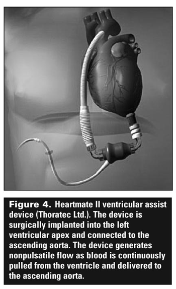
The average duration of mechanical support is 155 days. The current device utilized in BC is the Heartmate II (Thoratec Ltd., California). It generates continuous, nonpulsatile blood flow through a rotary impellar, thereby draining blood from the left ventricle to the ascending aorta (Figure 4).
Compared with its predecessors, which produced pulsatile flow, this device is smaller and requires less aggressive anticoagulation (INR 2 to 3) to prevent thromboembolic events.
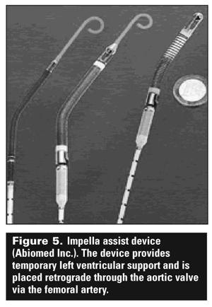
The Impella (Abiomed Inc., Massachusetts) is a temporary, percutaneously inserted assist device that is placed retrograde across the aortic valve into the left ventricle via the femoral artery (Figure 5). It also generates nonpulsatile flow via a rotary device. It is designed for short-term (7 to 10 days) support. Unlike an intra-aortic balloon pump it has no effect on afterload, and purely augments cardiac output.
Up to 50% of patients undergoing cardiac transplantation have pre-existing VAD support.11 Ventricular assist devices are currently approved as a bridge to transplant or recovery of the native heart in patients who are transplant candidates. Destination therapy (implantation of a VAD into a patient who is not a candidate for transplantation) is not yet approved; however, this remains an area of ongoing discussion.
Conclusions
Cardiac transplantation remains a life-saving therapy for patients with end-stage heart failure. Patients with NYHA class III or IV heart failure symptoms and no contraindications should be referred for evaluation. The assessment and management of these patients require a multidisciplinary team.
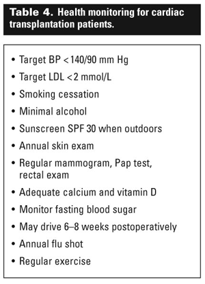
Standard malignancy screening and aggressive management of classic atherosclerotic risk factors are imperative for patients after transplantation (Table 4). The ongoing scarcity of available donor organs has led to the development of ventricular assist devices. These may allow patients to survive long enough to receive an organ. Alternatively, as these devices advance, there may come a day when mechanical support replaces the allograft.
Competing interests
None declared.
References
1. Taylor D, Edwards L, Aurora P, et al. Registry of the International Society for Heart and Lung Transplantation: Twenty-fifth official adult heart transplant report—2008. J Heart Lung Transplant 2008;27:943-956.
2. Haddad H, Isaac D, Legare JF, et al. Canadian Cardiovascular Society Consensus Conference update on cardiac transplantation 2008: Executive Summary. Can J Cardiol 2009;25:197-205.
3. Aaronson KD, Schwartz JS, Chen TM, et al. Development and prospective validation of a clinical index to predict survival in ambulatory patients referred for a cardiac transplant evaluation. Circulation 1997;95:2660-2667.
4. Ross H, Hendry P, Dipchand A, et al. 2001 Canadian Cardiovascular Society Consensus Conference on cardiac transplantation. Can J Cardiol 2003;19:620-654.
5. Betkowski AS, Graff R, Chenn JJ, et al. Panel-reactive antibody screening practices prior to heart transplantation. J Heart Lung Transplant 2002;21:644-650.
6. Itescu S, John R. Interactions between the recipient immune system and the left ventricular assist device surface: Immunological and clinical implications. Ann Thorac Surg 2003;75(6 suppl):S58-65.
7. Hertz MI, Aurora P, Christie JD, et al. Registry of the ISHLT: A quarter century of thoracic transplantation. J Heart Lung Transplant 2008;27:937-942.
8. Milpied N, Vasseur B, Parquet N, et al. Humanized anti-CD20 monoclonal antibody (Rituximab) in post transplant B-lymphoproliferative disorder: A retrospective analysis on 32 patients. Ann Oncol 2000;11:113-116.
9. Haddad M, Pflugfelder PW, Guiraudon C, et al. Angiographic, pathologic, and clinical relationships in coronary artery disease in cardiac allografts. J Heart Lung Transplant 2008;24:1218-1225.
10. Buszman P, Zembala M, Wojarski J, et al. Comparison of intravascular ultrasound and quantitative angiography for evaluation of coronary artery disease in the transplanted heart. Ann Transplant 1996;1:31-33.
11. Taylor DO, Edwards LB, Boucek MM, et al. Registry of the IHLST: Twenty-fourth official adult heart transplant report—2007. J Heart Lung Transplant 2007;26:769-781.
Dr Stadnick is a cardiology fellow at the University of British Columbia. Dr Ignaszewski is head of the University of British Columbia Division of Cardiology at Providence Health Care (St. Paul’s Hospital). He is also the acting medical director of the BC Transplant Heart Transplant Program.