Underestimation of life expectancy in elderly patients: The example of paraplegia
Issue: BCMJ,
vol. 45 , No. 4 , May 2003 ,
Pages 178-182 Clinical Articles
Detailed tables of life expectancy in patients with spinal cord injury have appeared in recent reports from the US (1995) and England (1998). For patients with paraplegia, both studies give estimates around 80% of normal at age 20, but by age 80 the estimates are down to 50% of normal in one study and 20% in the other. We show that all of the estimates at age 80 are too low, due to a downward bias in the conventional methods of calculation. An alternative approach is suggested, based on the trend in proportional life expectancy from younger ages. Support for this approach is provided by a recent recalculation of data from the US study.
Conventional estimates of life expectancy in elderly patients with life-shortening conditions are almost always lower than they should be. An alternative approach is suggested, based on the trend in proportional life expectancy from younger ages.
A pedestrian-automobile accident has left Dorothy with paraplegia at T10. Her future costs of care will be covered by the driver’s insurance, but there is disagreement over her life expectancy. Dorothy is currently 80 years old, at which age the average life expectancy for a female is around 9 years (Canada 9.3; England 8.4; US 9.1). Two experienced doctors have given their opinion on Dorothy’s life expectancy: one has suggested 4.2 years; the other 1.8 years. Each estimate was based on a different follow-up study, but both studies were of high quality so there was little reason to choose one over the other. The case has now been settled out of court, and a structured settlement (life annuity) has been purchased to ensure that Dorothy will receive regular payments for care until she dies, whatever her actual length of survival.
As part of the settlement, Dorothy’s life expectancy was assumed to be the average of the two expert opinions, that is, 3 years, but we present evidence that 6 years would be a more accurate figure. Because of the underestimation of her life expectancy, the annual funding for Dorothy’s care will now be approximately half what it should have been.
Doctors are often asked to estimate life expectancy in patients who have survived a serious accident or developed a life-threatening disease. Evidence from epidemiological studies can be very helpful, but actual estimates of life expectancy are rarely provided in published articles. Spinal cord injury has been a notable exception, with some life expectancy figures appearing in reports from a Canadian study as early as 1961 through to 1983.[1]
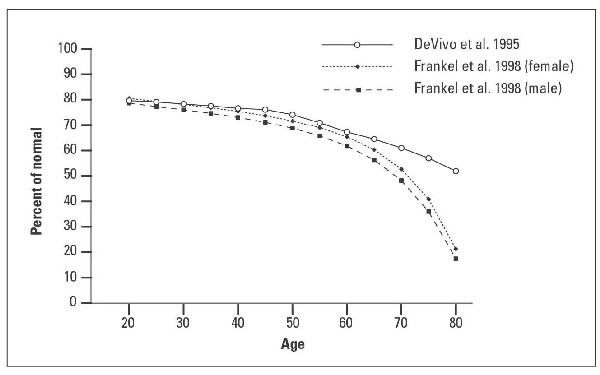
The first detailed table was published in 1995, when DeVivo and colleagues provided life expectancy estimates for four categories of spinal cord injury at 5-year intervals between ages 5 and 80.[2] The top line of Figure 1 shows the proportional (or “relative”) life expectancy estimates from this report for the category of paraplegia. Each point shows the life expectancy at that age as a percentage of the normal value at the same age. Thus at age 20 the estimated life expectancy of 44.8 years is 80% of the normal value of 56.3 years.
There is little change over the first 30 years, with a drop of only 6% by age 50 (to 74%; 21.2 years vs the normal figure of 28.6 years). The rate of decline then accelerates, so that over the next 30 years the percentage figure declines a further 22% by age 80 (to 52%; 4.2 years vs 8.1 years).
The second and third lines in Figure 1 show the comparable figures from a report by Frankel and colleagues published in 1998, which had the added feature of separate estimates for males and females.[3] The proportional life expectancy values are seen to be slightly higher in females than males at all ages, and both sets of figures are close to the DeVivo figures between ages 20 and 60. After age 60 the Frankel figures decline more rapidly, and by age 80 they are down to only 22% in females (1.8/8.3) and 17% in males (1.1/6.4).
The reason for these much lower estimates in the Frankel data by age 80 is not immediately obvious, since although the Frankel standardized mortality ratio (SMR) is higher in the oldest age group (see the Table: 3.68 vs 2.21) this alone cannot explain such a large difference between the two studies. This issue is explored further in later sections of this article, where the results from each study are examined in more detail.
The usual method of calculating life expectancy is by means of a life table that follows a hypothetical group of 100 000 newborn children, with current death rates being applied at each year of age. By about age 110 the last survivor has died, and the average life expectancy at each year of age can then be calculated at each age by dividing the remaining person-years of survival by the number alive at the start of the same year.
In persons with a life-shortening condition such as paraplegia, the life expectancy at each age can be obtained in the same way, except that the annual rates in the life table will need to be increased. This is usually done by multiplying the normal rates in the life table by one or more standardized mortality ratios derived from a suitable follow-up study, thus converting the normal expected rates of the life table to the observed rates seen in the study. A small follow-up study will often only supply a single SMR, which is then applied to all ages in the life table.[4] With larger studies there may be several age-specific SMRs available, based on age groups such as 20–24, 25–29, and so on.
Singer and others have suggested that in some situations a better method of changing the rates in the life table would be to add the difference between observed (O) and expected (E)—that is, adding O-E to the normal rate, rather than using the SMR (the ratio O/E) to multiply the rates.[5] In life expectancy studies this difference between observed and expected rates is usually referred to as the excess death rate (EDR).
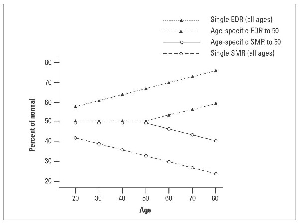
The effect of applying a single EDR to the life table is almost the exact opposite of a applying a single SMR. Thus when a single (constant) SMR is applied to the whole life table the proportional life expectancy declines with age, but with a constant EDR it increases with age. These trends are shown by the outer lines in the simplified schematic diagram of Figure 2.
The two inner lines of Figure 2 show what can occur when age-specific SMRs and EDRs are available below (say) age 50, leaving a final age group of 50 plus. The life expectancy estimates are now very similar until age 50, but the final age group now has a constant SMR or EDR from 50 to about 110, so the slopes again diverge and run parallel to the two outer lines.
Results of DeVivo and colleagues (1995)
This report is from the large US program of model care systems that began in 1973. By the time of the 1995 report, the study had recruited over 17000 persons with spinal cord injury, and follow-up was very good, with successful tracing of 92% of the deaths and 99% of the survivors.[2]
Age-specific standardized mortality ratios were calculated for four age groups, and these are shown in the Table, together with the estimated overall SMR for all ages combined.
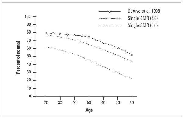
In Figure 3 the top line shows the published life expectancy estimates as a percentage of normal (as in Figure 1), while the second line shows the effect of multiplying all the rates in the life table by the single all-age SMR of 2.8.
The third line shows the result of doubling this figure to 5.6, to illustrate that the size of the SMR has little effect on the shape of the curve. Indeed, all three lines would be almost parallel if it were not for the initial flattening of the top line, due to the three age-specific SMRs at younger ages.
Note that this early flattening of the curve was also seen in the third line in Figure 2 (the schematic diagram) due to the age-specific SMRs below age 50, followed by a decline across the single age group beyond age 50. (No EDR values were provided in the DeVivo report, but as in the second line of Figure 2, if they had been available, the proportional life expectancy would eventually start rising, and would be approaching 90% by age 80.)
Results of Frankel and colleagues (1998)
The total follow-up period in this study was a remarkable 50 years, and again the follow-up was very good, with successful tracing of 92% of the subjects. The study was based mainly on patients who had been treated at the National Spinal Injuries Centre at Stoke Mandeville, with additional data from the Regional Centre at Southport.[3] Although the full report covered the years 1943–92, the authors restricted the estimates of life expectancy to 1973–90 to reflect recent mortality experience, and to allow for direct comparison with the 1995 data from DeVivo and colleagues.
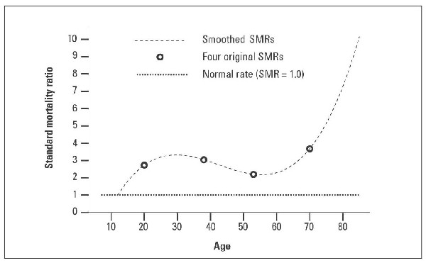
During the 1973–90 period a total of 1672 subjects were recruited, approximately 10% of the 17000 in the DeVivo study, so that division of the data into the same amount of detail led to some SMRs being based on relatively small numbers. To reduce variations attributable to the resulting instability of the SMRs, the authors explain that they applied smoothing techniques “using a third degree polynomial regression.”
Unfortunately, due to the distribution of SMR values in the paraplegia data, this polynomial equation generates very high SMRs at the older ages. This is illustrated in Figure 4, where the smooth S-shaped curve passes through each of the four original SMR values, but as it reaches the last SMR (at 3.68) the line is rising so steeply that the SMR has almost doubled (to 7) by age 80, and is approaching the ceiling value of 10 by age 85. (Here the normal death rate is approximately 10% per year, so simple multiplication by 10 would give 100%—that is, no survivors. This would account for the very steep decline in proportional life expectancy seen in Figure 1, where both male and female lines appear to be headed for zero at around age 85.)
Using the trend from younger ages
The rapid drop in life expectancy at the oldest ages in the Frankel study is the result of an unusual problem, in which the process of smoothing has created artificially high SMR values above age 70. In contrast, the underestimation of life expectancy in the DeVivo study is less severe, but it is a much more common problem, because in most cases only a single (constant) age-specific standardized mortality ratio is available for elderly subjects. The result is that the conventional practice of using a fixed SMR above a certain age will almost always lead to an unduly pessimistic estimate of life expectancy.
An alternative approach has recently been suggested, based on the observation that proportional life expectancy figures tend to remain constant with age, or show only a slight upward or downward trend. This pattern has been documented in several life-threatening conditions, and provides the most intuitively simple solution to what is otherwise a very difficult problem.[6]
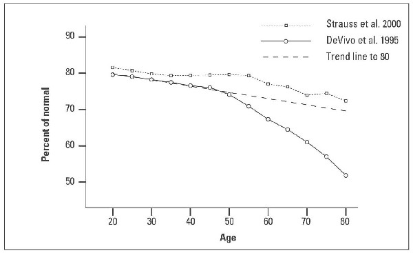
In the present case, this approach is illustrated in Figure 5, where the 20–50 trend line in the DeVivo data has been extended to age 80, at which point it reaches a value of 70%—equivalent to a life expectancy of approximately 6 years (Canada 6.5; England 5.9; US 6.4).
It is, of course, rarely possible to test the validity of this approach, because the information that is needed is itself the source of the problem, that is, most studies do not have enough elderly patients. However, the large size of the US model systems study has enabled Strauss and colleagues to calculate age-specific SMRs by 5-year age groups from age 10 through to age 80.[7] The results for paraplegia are shown by the top line in Figure 5, which is almost parallel to the DeVivo trend line, and reaches a value of 72% at age 80—virtually identical to the trend line prediction of 70%.
Since they were introduced in the 1980s, structured settlements have helped ensure that many patients receive steady funding for the rest of their lives. However, there is still a need for reasonably accurate estimations of individual life expectancy. Thus in the example of Dorothy, her structured settlement was purchased with a lump-sum award based on a predicted 3 years of care. It is therefore providing her with approximately half the annual amount that would have resulted from the more realistic life expectancy of 6 years.
With current methods, life expectancy in the elderly is underestimated when only a single SMR is applied to the whole life table, or whenever age-specific SMRs do not extend beyond the age of the patient. These errors are not confined to persons with spinal cord injury, but will occur in the presence of any life-shortening condition.
In the absence of reliable age-specific SMRs (or EDRs) for older age groups, the simple extrapolation of proportional life expectancy from younger ages will usually provide the best estimate of life expectancy in an elderly patient.
Table. Paraplegia: Age-specific standardized mortality ratios in two reports.
| Age | 0-30 | 31-45 | 46-60 | 61+ | All ages |
| DeVivo et al.[2]1995
Frankel et al.[3] 1998 |
3.86
3.14 |
3.74
3.04 |
1.96
2.20 |
2.21
3.68 |
(estimated) 2.80
2.83 |
We thank Ian Karp, Philip Steele, Keith Chambers, and Janet Heavyside for their helpful comments and suggestions.
None declared.
References
1. Geisler WO, Jousse AT, Wynne-Jones M, et al. Survival in traumatic spinal cord injury. Paraplegia 1983;21:364-373. PubMed Abstract
2. DeVivo MJ, Stover SL. Long-term survival and causes of death. In Spinal cord injury: Clinical Outcomes from the Model Systems. Gaithersburg, MD: Aspen Publishers, 1995:289-316. Full Text
3. Frankel HL, Coll JR, Charlifue SW, et al. Long term survival in spinal cord injury: A fifty year investigation. Spinal Cord 1998;36:266-274. Abstract Full Text
4. Smart CN, Sanders CR. The Costs of Motor Vehicle Related SCI. Washington, DC: Insurance Institute for Highway Safety, 1976.
5. Singer RB. A method of relating life expectancy in the US population life table to excess mortality. J Insur Med 1992;24:32-41. PubMed Citation
6. Anderson TW. Life Expectancy in Court. Vancouver, BC: Teviot Press, 2002.
7. Strauss D, DeVivo MJ, Shavelle R. Long-term mortality risk after spinal cord injury. J Insur Med 2000;32:11-16.
Terence W. Anderson BM, BCh, FRCPC, PhD and Stephen A. Marion, MD, FRCPC
Dr Anderson is professor emeritus in the Department of Health Care and Epidemiology, Faculty of Medicine, University of British Columbia. Dr Marion is a professor in the Department of Health Care and Epidemiology, Faculty of Medicine, UBC.
