
Serotonin syndrome presenting as posterior reversible encephalopathy syndrome in the setting of combined selective serotonin reuptake inhibitors and attention-deficit/hyperactivity disorder therapy
Issue: BCMJ,
vol. 68, No. 8, October 2026
|
Pages 280-284 |
Clinical Case Reports | doi.org/10.66794/2c69
ABSTRACT: Serotonin syndrome is a potentially life-threatening toxidrome resulting from excess serotonergic activity. Posterior reversible encephalopathy syndrome is a rare and serious complication of serotonin syndrome; there are only two reported cases in the literature. We present the case of a 38-year-old woman with a history of anxiety, managed on escitalopram, and a recent diagnosis of attention-deficit/hyperactivity disorder. She had been started on lisdexamfetamine dimesylate and dextroamphetamine prior to presentation. She subsequently developed serotonin syndrome following the addition of cyclobenzaprine for musculoskeletal pain. She presented with evidence of neurologic dysfunction, and neuroimaging demonstrated watershed ischemia and multifocal white matter changes consistent with posterior reversible encephalopathy syndrome. Extensive workup excluded alternative causes. Following discontinuation of serotonergic agents and supportive care, complete clinical and radiological resolution was achieved. This case highlights the risk of polypharmacy in patients with multiple psychiatric comorbidities and underscores the importance of vigilant medication reconciliation.
Clinicians managing patients with attention-deficit/hyperactivity disorder should be vigilant about the underrecognized risk factor of polypharmacy in the development of serotonin syndrome.
Serotonin syndrome is a potentially life-threatening drug-induced toxidrome associated with increased serotonergic activity in both the peripheral and central nervous systems.[1] It is characterized by a dose-dependent spectrum of clinical findings, including cognitive impairment, autonomic hyperactivity, and neuromuscular abnormalities.[1,2] The spectrum of cognitive dysfunction associated with serotonin syndrome ranges from mild irritability to profound coma, while hyper-reflexivity, clonus, and tremor represent a portion of the diverse neuromuscular abnormalities.[1,2] Autonomic manifestations include nausea, vomiting, diarrhea, dizziness, tachycardia, hypertension, mydriasis, hyperactive bowel sounds, diaphoresis, fever, and sexual dysfunction.[1,2] While hypertension is a common feature of serotonin syndrome, it typically falls within the mild to moderate range and does not necessitate specific therapy.[2] However, in some cases, severe dysautonomia can lead to hypertensive crisis, posterior reversible encephalopathy syndrome, myocardial infarction, and even death.[2,3]
Serotonin syndrome is a diagnosis of exclusion; there is no single diagnostic test that confirms the syndrome. However, clinical diagnosis can be determined based on the Hunter Serotonin Toxicity Criteria, which require the use of a serotonergic agent and one of the following: spontaneous clonus, inducible clonus plus agitation or diaphoresis, ocular clonus plus agitation or diaphoresis, tremor plus hyperreflexia, or hypertonia plus temperature above 38 °C plus ocular clonus or inducible clonus.[4] A summary of the features, diagnosis, and management of serotonin syndrome is provided in Box 1.
BOX 1. Key clinical features and management of serotonin syndrome.[1,2,4]
Definition
Serotonin syndrome is a potentially life-threatening drug reaction resulting from excess serotonergic activity in the central and peripheral nervous systems. It is characterized by a clinical triad of:
- Mental status changes (e.g., agitation, confusion, anxiety)
- Autonomic instability (e.g., tachycardia, hyperthermia, diaphoresis, labile blood pressure)
- Neuromuscular abnormalities (e.g., clonus, hyperreflexia, myoclonus, tremor)
Diagnosis: Hunter Serotonin Toxicity Criteria
In the setting of a serotonergic agent, one of the following must be present:
- Spontaneous clonus
- Inducible clonus + agitation or diaphoresis
- Ocular clonus + agitation or diaphoresis
- Tremor + hyperreflexia
- Hypertonia + temperature > 38 °C + ocular or inducible clonus
Severity classification
- Mild: Tremor, tachycardia, diaphoresis, mydriasis, and intermittent tremor or myoclonus
- Moderate: Mild symptoms + hyperreflexia, clonus, agitation, and hyperthermia (≤ 41 °C)
- Severe: Hyperthermia (> 41 °C), rhabdomyolysis, renal failure, seizures, and respiratory failure; can be fatal
Management
- Discontinue all serotonergic agents immediately
- Supportive care: IV fluids, oxygen, and cardiac monitoring
- Benzodiazepines: First-line treatment for agitation and neuromuscular hyperactivity
- Active cooling for hyperthermia; avoid antipyretics (ineffective for this mechanism)
- Severe cases: ICU admission, intubation, or neuromuscular paralysis if refractory
Over the past 2 decades, diagnoses of attention-deficit/hyperactivity disorder (ADHD) have risen steadily.[5] Although the cause is debated, clinicians are increasingly likely to encounter patients who are receiving treatment for ADHD. Notably, 65% to 80% of children and up to 85% of adults with ADHD have at least one psychiatric comorbidity, which significantly increases the risk of polypharmacy and drug interactions.[6,7] Stimulant medications, such as lisdexamfetamine dimesylate and dextroamphetamine, are commonly prescribed for the management of ADHD. These agents are thought to primarily enhance dopamine and norepinephrine signaling in the central nervous system.[8,9] However, they may also have indirect effects on serotonin levels. When used alone, stimulant medications are generally considered safe and effective.[8] However, caution is warranted when combining them with other medications that modulate serotonin function, because this may increase the risk of serotonin syndrome.[1]
Posterior reversible encephalopathy syndrome was first identified as a distinct neurological condition in 1996.[10] Over time, it has gained recognition as a compelling clinical and radiological syndrome. Among patients with posterior reversible encephalopathy syndrome, common symptoms encompass altered mental status, headache, seizures, visual disturbances, focal neurological deficits, and status epilepticus.[11] Posterior reversible encephalopathy syndrome is now a more recognized condition in some patients hospitalized for acute illness.[11,12] This syndrome, first described as a possible consequence of hypertension, eclampsia, or immunosuppressive therapy, has more recently been recognized in a variety of disorders, including several of iatrogenic or neurotoxic etiology.[10,11] Despite its recognized clinical features, the precise pathogenesis of posterior reversible encephalopathy syndrome remains elusive, although it is believed to be associated with the loss of cerebral autoregulation and endothelial dysfunction.[13] To our knowledge, there have been only two other reported cases of serotonin syndrome leading to posterior reversible encephalopathy syndrome.[14,15]
Posterior reversible encephalopathy syndrome is diagnosed based on clinical and radiological assessment. Neuroradiological imaging usually shows edema involving the cerebral posterior regions.[11,12,16,17] Three common imaging patterns seen with posterior reversible encephalopathy syndrome are a dominant parieto-occipital pattern, a superior frontal sulcus pattern, or a holo-hemispheric watershed pattern.[16] The diagnosis is usually made based on compatible clinical and radiological patterns.[3] However, when the diagnosis is uncertain, the clinical and radiological improvement that occurs with appropriate treatment supports the diagnosis.[11] Treatment of posterior reversible encephalopathy syndrome includes correcting elevated blood pressure, discontinuing causative agents, initiating anti-epileptic therapy if seizures are present, and addressing the underlying causes.[11-13] The overall prognosis for posterior reversible encephalopathy syndrome is favorable with early diagnosis and treatment, because the symptoms and imaging findings can reverse after promptly discontinuing causative agents and administering appropriate treatment.[3]
In this case report, we describe a patient who initially presented with nonspecific neurological symptoms. She had a preliminary diagnosis of serotonin syndrome, and her imaging findings were consistent with posterior reversible encephalopathy syndrome.
Case data
A 38-year-old woman with a history of anxiety, ADHD, and polysubstance use disorder presented with nonspecific neurological symptoms to the emergency department in a hospital in rural British Columbia. She presented after feeling unwell for approximately 1 week. Initially, she noted nausea and abdominal pain. Four days prior to presentation, she was started on indomethacin 25 to 50 mg three times a day as needed and cyclobenzaprine 10 mg daily for shoulder pain. On the morning of her presentation, she noted that her hands seemed to be slowed, as though they were not cooperating. Later that day, she developed a frontal headache with visual disturbances, lightheadedness, and unsteadiness.
In the 3 months prior, this patient was diagnosed with ADHD and started on stimulant medications, which included lisdexamfetamine dimesylate 60 mg daily and dextroamphetamine 5 to 10 mg daily as needed. She also took escitalopram daily. Although she had a history of substance use, she had not used any substances in 1 year.
Initial physical examination revealed a heart rate of 94 bpm, blood pressure of 107/73, temperature of 37 °C, and oxygen saturation of 99% on room air. Her general examination did not show any evidence of nuchal rigidity, lymphadenopathy, or skin rashes. There were no cardiac murmurs or abnormalities. Her neurological examination showed an alert and oriented female who appeared agitated. Her cranial nerves were intact, with no nystagmus. She had no sensory deficits or neglect and no motor drift. She was noted to have spontaneous choreiform-type movement in both of her upper limbs. She demonstrated bilateral dysmetria and bilateral dysdiadochokinesis. Hyperreflexia was noted in all limbs, and four beats of clonus were noted in both feet. No tremor was observed.
Initial lab tests were significant for an elevated white blood cell count at 15 × 109 cells/L, a mild C-reactive protein elevation of 45 mg/L, and a troponin elevation of 3553 ng/L. Urinalysis was within normal limits. Urine toxicology screen was positive for tricyclic antidepressants and amphetamines. The patient’s initial CT head with angiography did not show any large vascular abnormality or occlusion. Crowding of the lateral ventricles was reported and suggested possible increased intracranial pressure. Given the history of using multiple serotonergic agents, recent medication change, and clinical features that fulfilled the Hunter Serotonin Toxicity Criteria, the patient was suspected of having serotonin syndrome.[4]
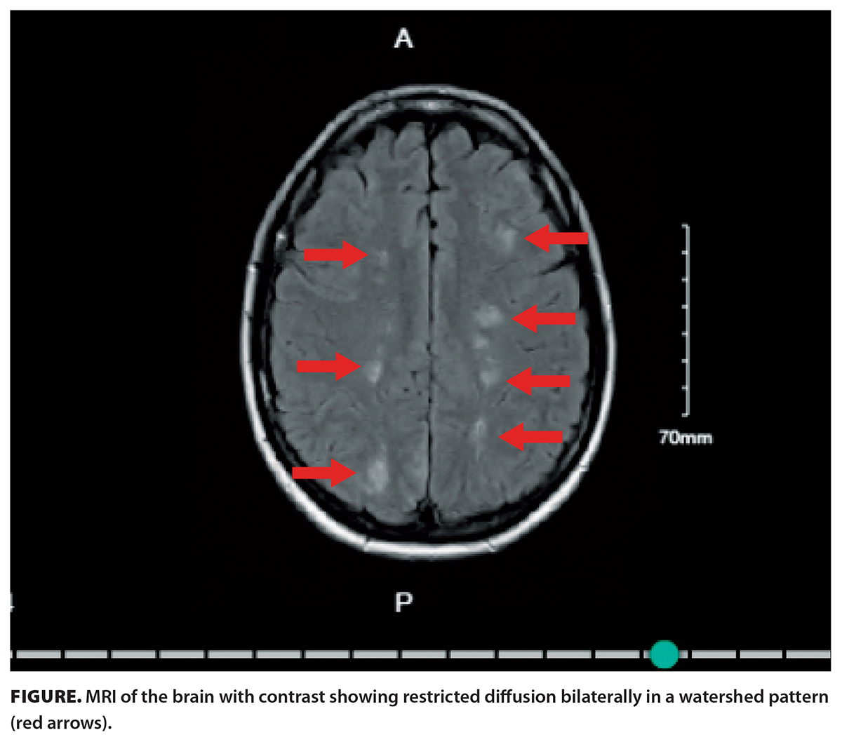 The initial CT angiogram head findings, along with the patient’s physical examination abnormalities, prompted a noncontrast head MRI to be performed. The diffusion-weighted sequences showed small multicentric areas of recent ischemia in a watershed distribution between the middle cerebral artery and anterior cerebral artery territory and involving the posterior occipital lobes, suggesting peripheral posterior cerebral artery territory involvement [Figure].
The differential diagnosis for these MRI findings in the context of the patient’s presentation was broad, including stroke, vasculitis, and infection. Further investigations, including full cerebrospinal fluid analysis; rheumatologic workup including antinuclear antibody, rheumatoid factor, and antineutrophil cytoplasmic antibodies; and an extended infectious workup including HIV, Lyme disease, and treponemal antibodies, all returned negative. A transthoracic echocardiogram was unremarkable, and no significant valvular dysfunction was noted. A bubble study was also negative. Bilateral leg ultrasound with Doppler did not reveal any deep vein thromboses; an antiphospholipid antibody syndrome workup was also negative.
A contrast MRI was performed the following day, which again showed symmetric bilateral scattered multifocal hyperintense lesions throughout the subcortical white matter. In discussion with neurology and in the setting of a negative workup for other causes, the diagnosis of posterior reversible encephalopathy syndrome was proposed. In this clinical case, it was suspected that posterior reversible encephalopathy syndrome was a result of the patient’s initial diagnosis of serotonin syndrome.
Upon admission, all serotonergic agents were discontinued, and the extensive investigations outlined above were ordered. The patient was initially treated with antibiotics, acyclovir, and aspirin. Once her cerebrospinal fluid was analyzed and did not suggest infection, her antimicrobials were discontinued. Over the following 2 weeks, she was treated supportively with intensive physical therapy. The patient’s neurological symptoms slowly improved during her stay. At the time of discharge, she self-reported some cognitive difficulties and confusion, as well as ongoing visual disturbance, but her gait had improved. She was seen in the community 8 weeks after discharge and had complete resolution of all her symptoms. Her repeat MRI at this time showed complete resolution of all cortical foci. After discussion in the clinic, she elected to remain on aspirin 81 mg daily indefinitely.
Discussion
Serotonin syndrome is a potentially life-threatening toxidrome arising from excess serotonergic activity in the central and peripheral nervous systems.[2] It most commonly results from combinations of serotonergic agents that increase synaptic serotonin through complementary mechanisms, including increased synthesis, impaired reuptake, and direct receptor agonism that results in overstimulation of the 5-HT1A and 5-HT2A receptors.[2] The clinical presentation exists on a dose-dependent spectrum encompassing cognitive dysfunction, autonomic instability, and neuromuscular hyperactivity, with diagnosis typically established through the Hunter Serotonin Toxicity Criteria [Box 1].[1,2,4] Autonomic dysregulation in serotonin syndrome reflects exaggerated sympathetic outflow, most commonly manifesting as mild to moderate hypertension that seldom necessitates targeted intervention.[2,3] However, in severe cases, catecholamine surge can precipitate hypertensive crisis with end-organ consequences, including myocardial infarction, acute kidney injury, and, in rare instances, posterior reversible encephalopathy syndrome or death.[2,3]
Posterior reversible encephalopathy syndrome is diagnosed based on clinical and radiological assessment. Clinically, it is characterized by transient neurological symptoms, including headache, altered mental status, visual disturbances, focal deficits, and seizures.[3,11] MRI remains the neuroimaging modality of choice, with T2-weighted sequences demonstrating greater diagnostic sensitivity than CT, classically revealing vasogenic edema predominantly involving the posterior cerebral regions.[11] Once considered a sequela of hypertension alone, posterior reversible encephalopathy syndrome is now recognized across a broader spectrum of precipitating conditions, including drug-induced syndromes [Box 2].[3,11] The exact pathophysiology remains unclear, but it is believed that under normal physiological conditions, cerebral autoregulation maintains stable perfusion across a range of systemic pressures. It is hypothesized that in posterior reversible encephalopathy syndrome, this mechanism is overwhelmed or disrupted, leading to forced hyperperfusion, breakdown of the blood–brain barrier, and resultant vasogenic edema.[3,11] Endothelial dysfunction, whether driven by hypertension, circulating toxins, or inflammatory mediators, is believed to act synergistically with autoregulatory failure.[3] A distinctive characteristic of posterior reversible encephalopathy syndrome is the reversibility of clinical and radiological abnormalities after appropriate treatment and removal of precipitating factors.[3,11]
BOX 2. Drugs and medical conditions associated with posterior reversible encephalopathy syndrome (PRES).[3]
Drugs associated with PRES
Oncologic
- Alkylating agents (e.g., cisplatin, ifosfamide)
- Antimetabolites (e.g., methotrexate, fluorouracil)
- Kinase inhibitors (e.g., sunitinib, ponatinib)
- Anti–vascular endothelial growth factor medications (e.g., bevacizumab, aflibercept)
- Proteosome inhibitors (e.g., bortezomib, carfilzomib)
- Hormone-modulating agents (e.g., enzalutamide)
- Chemotherapy agents (e.g., paclitaxel, vinblastine)
Miscellaneous
- Ondansetron
- Lorazepam
- Levetiracetam
- Methadone
- Lithium
- Propofol
- Phenytoin
Anti-infectious
- Hydroxychloroquine
- Voriconazole
- Acyclovir
- Linezolid
- Vancomycin
Immunosuppressants
- Calcineurin inhibitors (e.g., cyclosporin, tacrolimus)
- Corticosteroids (e.g., prednisone, dexamethasone)
- Monoclonal antibodies (e.g., ustekinumab)
- Human immunoglobulins
- Sphingosine 1-phosphate receptor modulators
Other immune modulators
- Azathioprine
- Mycophenolate
- Thalidomide
Medical conditions associated with PRES
- Uncontrolled hypertension
- Systemic lupus erythematosus
- Cryoglobulinemia
- Hemolytic uremic syndrome
- Hematopoietic stem cell transplantation
- Renal disease
- Eclampsia
- Sepsis
- Thrombotic thrombocytopenic purpura
- Solid organ transplantation
- Collagen vascular disease
- Other autoimmune diseases
Because ADHD diagnoses have risen steadily over the past 2 decades, clinicians are increasingly likely to encounter patients who require pharmacologic management of both ADHD and comorbid psychiatric conditions.[5] Stimulant medications such as lisdexamfetamine dimesylate and dextroamphetamine are among the most commonly prescribed agents for ADHD; they work primarily by enhancing dopamine and norepinephrine signaling in the central nervous system and indirectly stimulate the serotonin pathway.[8,9] When combined with other serotonergic medications, such as selective serotonin reuptake inhibitors, serotonin and norepinephrine reuptake inhibitors, or monoamine oxidase inhibitors, stimulants may act synergistically on the serotonin pathway, resulting in an additive or potentiating effect that substantially elevates the risk of serotonin toxicity.[8,9] This is particularly important to consider when such a high percentage of patients being treated for ADHD have additional comorbid psychiatric conditions in which most treatment options are serotonergic agents.[6]
We believe that in our case, the use of escitalopram, combined with the recent diagnosis and treatment of ADHD with both short-acting and long-acting stimulants, lowered this patient’s threshold for serotonin syndrome. When prescribed cyclobenzaprine for her musculoskeletal injury, this was enough to tip the balance, leading to the development of serotonin syndrome, which then resulted in posterior reversible encephalopathy syndrome, which presented with neurologic dysfunction and watershed infarcts on MRI.[18] In this case, the diagnosis of serotonin syndrome was based on the Hunter Serotonin Toxicity Criteria and the history of serotonergic medication use.[4] The patient remained normotensive throughout her admission; therefore, we believe that posterior reversible encephalopathy syndrome was not a result of hypertension from serotonin syndrome but rather was secondary to autoregulatory dysfunction from serotonin syndrome. In addition, an extensive list of alternative diagnoses was considered and ruled out, which further supported the diagnosis of serotonin syndrome resulting in posterior reversible encephalopathy syndrome. Moreover, a prompt resolution of both symptoms and imaging findings with cessation of serotonergic medications further supports a diagnosis of posterior reversible encephalopathy syndrome from serotonin syndrome and excludes other secondary possibilities, because a resolution of other pathologies would not be so immediate.
To our knowledge, this is one of the first cases of this combination of escitalopram, lisdexamfetamine dimesylate/dextroamphetamine, and cyclobenzaprine ultimately resulting in posterior reversible encephalopathy syndrome. This case highlights important principles for clinical practice. Clinicians who are managing patients with ADHD and prescribing stimulant medications should be vigilant about potential drug interactions, especially when combined with other agents that act on serotonergic pathways, such as escitalopram and cyclobenzaprine.
Summary
This case highlights important principles and the risk of polypharmacy for clinicians managing patients with ADHD and associated comorbidities. In particular, ADHD therapies are an underrecognized risk factor for the development of serotonin syndrome. This is especially relevant currently, because an increasing number of patients globally are being treated for ADHD, and many of them have additional psychiatric comorbidities. This case emphasizes the importance of a thorough medication history and awareness of potential adverse effects to prevent life-threatening complications when prescribing these medications given the increasing prevalence of polypharmacy in psychiatric and neurological disorders.
Competing interests
None declared.
This article has been peer reviewed.

This work is licensed under a Creative Commons Attribution-NonCommercial-NoDerivatives 4.0 International License.
References
1. Boyer EW, Shannon M. The serotonin syndrome. N Engl J Med 2005;352:1112-1120. https://doi.org/10.1056/NEJMra041867.
2. Scotton WJ, Hill LJ, Williams AC, Barnes NM. Serotonin syndrome: Pathophysiology, clinical features, management, and potential future directions. Int J Tryptophan Res 2019;12:1178646919873925. https://doi.org/10.1177/1178646919873925.
3. Fugate JE, Rabinstein AA. Posterior reversible encephalopathy syndrome: Clinical and radiological manifestations, pathophysiology, and outstanding questions. Lancet Neurol 2015;14:914-925. https://doi.org/10.1016/S1474-4422(15)00111-8.
4. Dunkley EJC, Isbister GK, Sibbritt D, et al. The Hunter Serotonin Toxicity Criteria: Simple and accurate diagnostic decision rules for serotonin toxicity. QJM 2003;96:635-642. https://doi.org/10.1093/qjmed/hcg109.
5. Abdelnour E, Jansen MO, Gold JA. ADHD diagnostic trends: Increased recognition or overdiagnosis? Mo Med 2022;119:467-473.
6. Faraone SV, Banaschewski T, Coghill D, et al. The World Federation of ADHD international consensus statement: 208 evidence-based conclusions about the disorder. Neurosci Biobehav Rev 2021;128:789-818. https://doi.org/10.1016/j.neubiorev.2021.01.022.
7. Espinet SD, Graziosi G, Toplak ME, et al. A review of Canadian diagnosed ADHD prevalence and incidence estimates published in the past decade. Brain Sci 2022;12:1051. https://doi.org/10.3390/brainsci12081051.
8. Docherty JR, Alsufyani HA. Pharmacology of drugs used as stimulants. J Clin Pharmacol 2021;61(Suppl 2):S53-S69. https://doi.org/10.1002/jcph.1918.
9. Oades RD. Dopamine–serotonin interactions in attention-deficit hyperactivity disorder (ADHD). Prog Brain Res 2008;172:543-565. https://doi.org/10.1016/S0079-6123(08)00926-6.
10. Fugate JE, Claassen DO, Cloft HJ, et al. Posterior reversible encephalopathy syndrome: Associated clinical and radiologic findings. Mayo Clin Proc 2010;85:427-432. https://doi.org/10.4065/mcp.2009.0590.
11. Bartynski WS. Posterior reversible encephalopathy syndrome, part 1: Fundamental imaging and clinical features. AJNR 2008;29:1036-1042. https://doi.org/10.3174/ajnr.A0928.
12. Hinchey J, Chaves C, Appignani B, et al. A reversible posterior leukoencephalopathy syndrome. N Engl J Med 1996;334:494-500. https://doi.org/10.1056/NEJM199602223340803.
13. Marra A, Vargas M, Striano P, et al. Posterior reversible encephalopathy syndrome: The endothelial hypotheses. Med Hypotheses 2014;82:619-622. https://doi.org/10.1016/j.mehy.2014.02.022.
14. Prakash S, Rathore C, Kaushikkumar R. Posterior reversible encephalopathy syndrome in a patient with serotonin syndrome. BMJ Case Rep 2020;13:e236450. https://doi.org/10.1136/bcr-2020-236450.
15. Malik MT, Majeed MF, Zand R. Serotonin syndrome presenting as a posterior reversible encephalopathy syndrome. Case Rep Neurol 2020;12:63-68. https://doi.org/10.1159/000505907.
16. Bartynski WS, Boardman JF. Distinct imaging patterns and lesion distribution in posterior reversible encephalopathy syndrome. AJNR 2007;28:1320-1327. https://doi.org/10.3174/ajnr.A0549.
17. Covarrubias DJ, Luetmer PH, Campeau NG. Posterior reversible encephalopathy syndrome: Prognostic utility of quantitative diffusion-weighted MR images. AJNR 2002;23:1038-1048.
18. Bansal V, Aranke M, Vu P, Javed S. Serotonin syndrome from combination hydrocodone and cyclobenzaprine in a patient with cerebral palsy. Pain Manag 2023;13:329-334. https://doi.org/10.2217/pmt-2023-0044.
Dr Hawe is an internal medicine resident at the University of British Columbia. Dr Wik is a staff general internist at East Kootenay Regional Hospital in Cranbrook, BC, and is affiliated with the UBC Faculty of Medicine, Division of General Internal Medicine.
Corresponding author: Dr Nicole Hawe, nhawe@student.ubc.ca.
Thank you for this fascinating case that I am sure will stimulate readers to think about the cumulative load of serotonin their patients on multiple medications are facing! It is a topic that prescribers are not well educated in and only something I began to appreciate and look for after a few years of seeing impressive polypharmacy in frail elders and in palliative care patients.
I wanted to note that the newer term "toxicity" is preferred by some, including me, for a couple of reasons. Mostly this is academic but the one clinically-relevant justification is that labelling it a "toxicity" helps convey that the impact/symptoms exists on a spectrum (rather than there being a discrete entity or threshold at which one 'tips over' into this syndrome). The connotation of "toxicity" is that the effect is cumulative, worse with more agents that work on serotonin, whereas with "syndrome" the dose-related aspect is not implied.
I wonder if when you note that the addition of cyclobenzaprine "tipped the balance" into serotonin syndrome, if the patient may have had more subtle signs of serotonin toxicity already? Perhaps the additional agent then "tipped" her into meeting the Hunter Serotonin Toxicity Criteria (we notice it because there is clonus, hyperthermia, and autonomic instability), but perhaps there were some symptoms before this; eg. was the nausea from the indocid or the serotonin load? did she have any tremor, cognitive changes, or anxious feelings before the cyclobenzaprine?
Subtle serotonin toxicity can cause difficulty for patients and is a real diagnostic challenge, especially if we don't think to include it on the differential. If clinicians expect to always see the key symptoms that make up a 'syndrome' we may underdiagnose, and patients with mild confusion and restlessness may be 'dismissed' (or worse) given more psychoactive medications, when deprescribing would be the safest and effective treatment.
What a great case and learning for all of us. Thank you again for putting in the time and effort to publish this.
(Jessica Otte, UBC Therapeutics Initiative)