
Pancreatic cystic lesions in adults: Diagnosis and management
Issue: BCMJ,
vol. 68, No. 9, November 2026
|
Pages 320-326 |
Review Articles
ABSTRACT: Pancreatic cystic lesions are being increasingly detected with cross-sectional imaging. Although most are benign, a subset carry malignant potential. Accurate classification into nonneoplastic and neoplastic subtypes is essential, because lesions such as intraductal papillary mucinous neoplasms and mucinous cystic neoplasms warrant closer monitoring or surgical evaluation. Diagnosis is guided by cross-sectional imaging. Magnetic resonance cholangiopancreatography is the preferred modality; endoscopic ultrasonography is reserved for indeterminate or higher-risk lesions. Major society guidelines use high-risk and worrisome features to direct referral, intervention, or surveillance, although thresholds vary. Most asymptomatic cysts without concerning features can be safely followed with interval imaging, whereas lesions with mural nodules, ductal dilation, or suspicious cytology require specialist assessment. Early recognition, risk stratification, and coordinated surveillance are critical to enable timely detection of malignancy. This review focuses on the adult population, in whom pancreatic cystic lesions are most prevalent.
Early recognition, risk stratification, and coordinated surveillance are critical to the timely detection of malignant pancreatic cystic lesions.
Pancreatic cystic lesions are being increasingly encountered in clinical practice due to the increased use of cross-sectional imaging.[1] Incidental pancreatic cysts are identified in up to 20% of patients undergoing abdominal MRI, and prevalence increases to up to 40% with age (particularly in those over age 70) and underlying comorbidities, such as diabetes or chronic pancreatitis.[2,3]
While most pancreatic cystic lesions are benign and asymptomatic, a clinically important subset represents premalignant or malignant neoplasms. The incidental discovery of a pancreatic cyst often raises concerns for both clinicians and patients, particularly given the high mortality rate associated with pancreatic adenocarcinoma. However, most lesions follow an indolent course, and distinguishing between low- and high-risk cysts is critical to avoid unnecessary interventions while ensuring timely cancer detection. Early identification of cyst type guides diagnostic testing, informs malignancy risk, and determines the need for specialist referral. Clinical decision making, however, is complicated by the variability among major society guidelines, which differ in their definitions of high-risk features, indications for further workup, and recommended surveillance intervals.[2-6]
In this context, primary care physicians play a pivotal role in the initial identification and interpretation of radiologic findings, as well as in counseling patients, initiating appropriate surveillance, and facilitating specialist referral when warranted. This review provides a practical, evidence-based overview of pancreatic cystic lesions for primary care. It focuses on classification, natural history, and current guideline recommendations for workup, surveillance, and referral.
This review focuses specifically on the diagnosis and management of pancreatic cystic lesions in adults, the population in which these lesions are overwhelmingly prevalent. Pancreatic cystic lesions are rare in children and typically differ in etiology (e.g., congenital, hereditary, or syndromic causes). When a cyst is identified incidentally in a child, magnetic resonance cholangiopancreatography (MRCP) is the imaging test of choice. It can be ordered as a dedicated pediatric pancreatic-protocol MRI/MRCP, with referral to pediatric gastroenterology or surgery given the differing management pathways in this age group.
 Classification
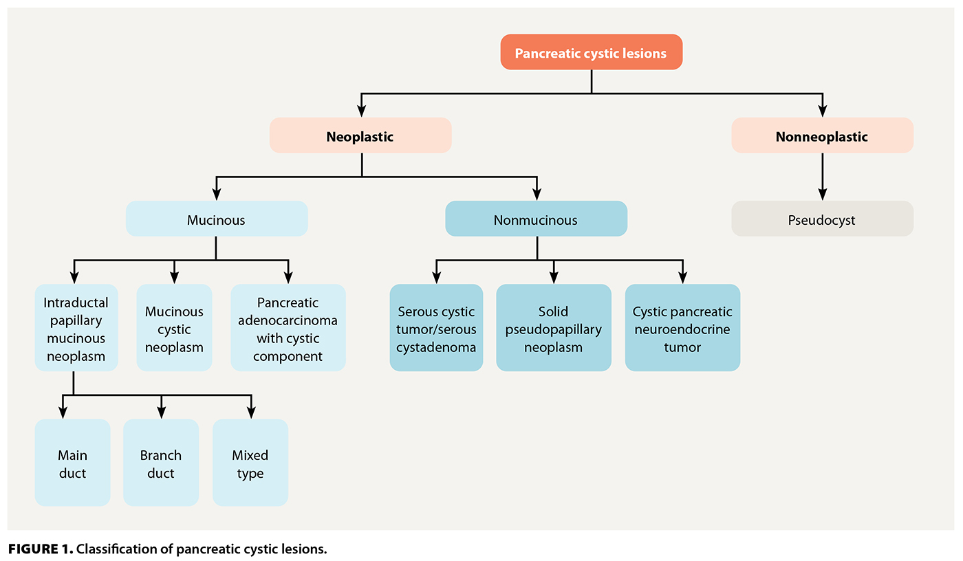
Pancreatic cystic lesions represent a heterogeneous group of entities with varying causes, epithelial linings, mucin production, and, most importantly, malignant potential. Broadly, they can be categorized into nonneoplastic and neoplastic lesions [Figure 1]. This classification is clinically important, because the risk of malignancy varies markedly among subtypes, and appropriate categorization guides diagnostic workup, referral, and surveillance strategies.
Nonneoplastic
Most nonneoplastic lesions are pseudocysts that represent collections of pancreatic secretions that have extravasated from a duct disrupted by inflammation or obstruction.[7] They typically occur in patients with a history of acute or chronic pancreatitis or abdominal trauma.
Other benign and uncommon cyst types include simple (true epithelial), lymphoepithelial, and retention cysts. Simple cysts are unilocular, are lined by a single layer of epithelium, lack communication with the pancreatic ductal system, and carry no malignant potential.[8] Lymphoepithelial cysts typically occur in middle-aged men; they can arise anywhere in the pancreas and are often multilocular, with a mean size of approximately 5 cm. They are benign but may mimic pseudocysts on imaging.[9,10] Retention cysts represent ductal side-branch dilation secondary to obstruction (e.g., calculi, mucin). They may have mucinous lining and can resemble intraductal papillary mucinous neoplasms, although they lack neoplastic potential.[11]
Neoplastic
Neoplastic pancreatic cystic lesions encompass a diverse group of cyst-forming tumors with varying malignant potential. The main subtypes are mucinous cystic neoplasms, intraductal papillary mucinous neoplasms, solid pseudopapillary neoplasms, serous cystic neoplasms/serous cystadenomas, cystic pancreatic neuroendocrine tumors, and cystic pancreatic adenocarcinoma.
Mucinous cystic neoplasms
Mucinous cystic neoplasms are mucin-producing epithelial cysts that occur in the body and tail of the pancreas, and they do not communicate with the ductal system. They occur almost exclusively in middle-aged women and are characterized histologically by ovarian-type stroma. Recent meta-analyses have reported an overall malignancy rate of 17% in resected specimens.[12]
Intraductal papillary mucinous neoplasms
Intraductal papillary mucinous neoplasms (IPMNs) are mucin-producing neoplasms that arise from the ductal epithelium. They are subclassified into three main types: main duct, branch duct, and mixed type. Malignant potential differs significantly by type. Main-duct IPMNs have a 30% to 50% risk of malignancy, whereas branch-duct IPMNs have a 3% to 12% risk.[13] IPMNs have ductal communication by definition and are a major cause of both referral and surveillance uncertainty.
Solid pseudopapillary neoplasms
Solid pseudopapillary neoplasms are large, well-encapsulated masses with mixed solid and cystic components, and they primarily affect young women.[14] Their malignant potential has not been fully characterized, but case series have reported malignancy in up to 15% of resected lesions. Local invasion or metastasis is possible, so surgical resection is indicated.[14]
Serous cystic neoplasms/serous cystadenomas
Serous cystadenomas are nonmucinous, glycogen-rich epithelial tumors with a classic honeycomb, microcystic appearance on imaging. They are essentially benign and have negligible risk of malignancy.[15] Cyst fluid analysis demonstrating von Hippel-Lindau gene alterations and the presence of vascular endothelial growth factor-A support a diagnosis of serous cystadenoma, although this is not routinely required.
Cystic pancreatic neuroendocrine neoplasms
Cystic pancreatic neuroendocrine neoplasms may exhibit secondary cystic degeneration that often appears as a well-circumscribed, hypervascular cystic mass, typically in the body or tail of the pancreas.[2] Malignant potential varies by grade and proliferative index, and management is individualized.
Pancreatic ductal adenocarcinoma with a cystic component
A subset of pancreatic ductal adenocarcinomas may undergo cystic degeneration or arise within a pre-existing cystic neoplasm, such as an IPMN or mucinous cystic neoplasm. They typically present as irregular, hypoechoic masses with an abrupt main-duct cutoff and upstream dilation and represent overtly malignant lesions that require oncologic resection when feasible.[2,16]
Key points: Classification
- Pancreatic cystic lesions are broadly divided into nonneoplastic (e.g., pseudocysts) and neoplastic lesions. Neoplastic cysts are further subdivided into mucinous (mucinous cystic neoplasm, intraductal papillary mucinous neoplasm [IPMN]) and nonmucinous (solid pseudopapillary neoplasm, serous cystic neoplasm/serous cystadenoma, cystic pancreatic neuroendocrine tumor, cystic pancreatic adenocarcinoma) subtypes.
- Malignant potential varies by subtype, from negligible in serous cystadenomas to 30% to 50% in main-duct IPMN; therefore, accurate classification is essential to guide management.
- Mucin-producing lesions (mucinous cystic neoplasm, IPMN) account for most clinically significant pancreatic cystic lesions and require the greatest attention given their premalignant potential.
Diagnosis
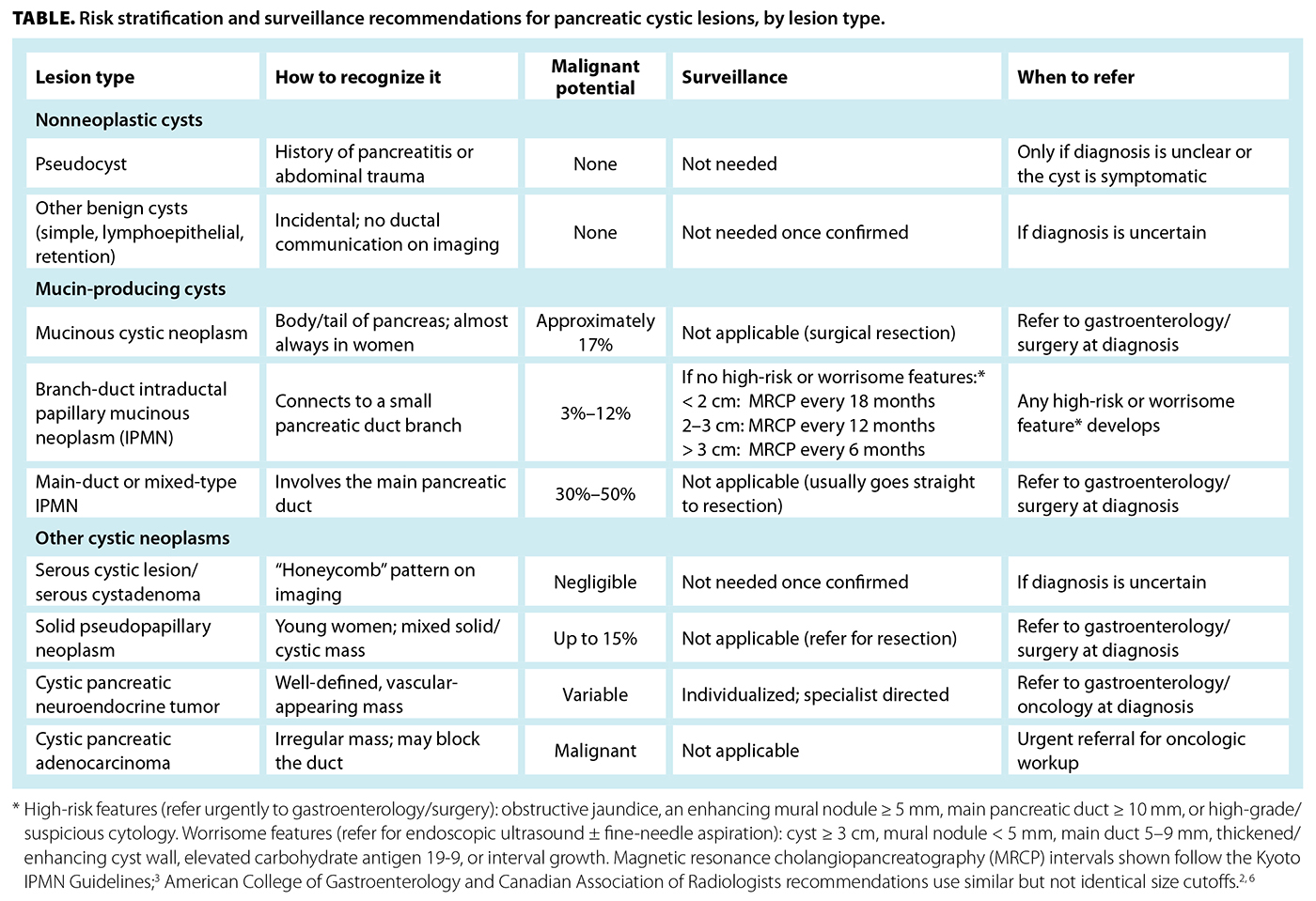
When a pancreatic cyst is discovered on cross-sectional imaging, the initial step is to confirm that it is a true cystic lesion and not an inflammatory fluid collection such as a pseudocyst. Reviewing the clinical history and any prior imaging is essential to assess chronicity and exclude pancreatitis-related causes. The subsequent diagnostic pathway is guided by lesion size, ductal communication, and presence of worrisome features [Table].
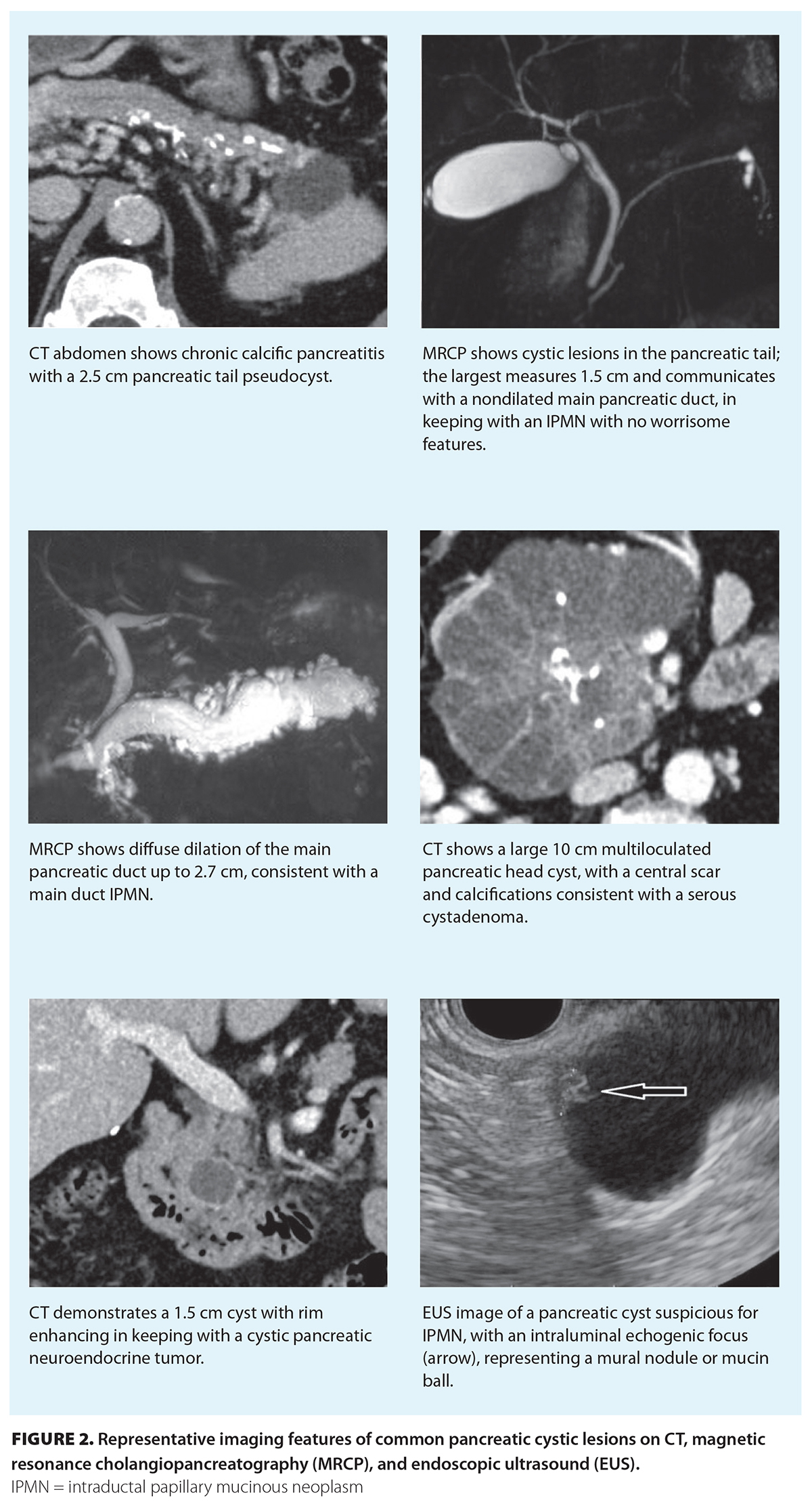 If the lesion is a true cyst, the next step is dedicated MRCP, which is the preferred modality for characterization. MRCP delineates ductal communication, defines the presence of mural nodules or septations, and identifies high-risk or worrisome features with superior sensitivity compared with CT.[2-4] When MRCP is contraindicated or nondiagnostic, a pancreatic-protocol contrast-enhanced CT is an acceptable alternative; it provides information about calcifications, wall thickening, and ductal dilation.[4,5] Representative cross-sectional and endoscopic imaging findings of common pancreatic cystic lesions are shown in Figure 2.
For lesions with indeterminate imaging features or worrisome characteristics, endoscopic ultrasonography offers superior spatial resolution and allows for fine-needle aspiration for cyst-fluid analysis. Cytology may reveal mucinous epithelium, cuboidal glycogen-rich epithelium suggestive of serous cystadenomas, high-grade dysplasia, or even malignant cells. Cyst-fluid biomarkers can further help refine diagnosis. Carcinoembryonic antigen greater than 192 ng/mL suggests a mucinous cyst, whereas less than 5 ng/mL supports a serous cyst. Glucose less than 2.8 mmol/L favors a mucinous lesion, while amylase is typically elevated if the cyst communicates with the ductal system.[2,16-18]
Despite these advances, surgical pathology remains the diagnostic gold standard when resection is undertaken because it allows for definitive histologic subtyping and grading of dysplasia.[19]
Key points: Diagnosis
- The initial evaluation should confirm a true pancreatic cyst and exclude inflammatory fluid collections, such as pseudocysts, based on clinical history and prior imaging.
- Magnetic resonance cholangiopancreatography is the preferred imaging modality for cyst characterization, including assessment of ductal communication and high-risk features.
- Endoscopic ultrasonography with fine-needle aspiration should be reserved for indeterminate or worrisome lesions, using cytology and cyst-fluid biomarkers (carcinoembryonic antigen, glucose, amylase) to refine diagnosis.
Risk stratification and surveillance
Risk stratification is central to determining whether a pancreatic cystic lesion requires surgical evaluation, endoscopic assessment, or continued surveillance. Major international guidelines have similar core criteria but differ in diagnostic thresholds and management aggressiveness. In general, lesions are evaluated for the presence of high-risk features, which warrant referral for surgical consideration, or worrisome features, which prompt further evaluation with endoscopic ultrasonography or short-interval imaging.
High-risk features indicate an increased risk of malignancy and generally prompt referral for further evaluation. These features include obstructive jaundice related to the cyst, an enhancing mural nodule 5 mm or larger, main pancreatic duct dilation 10 mm or larger, and cytology showing high-grade dysplasia or carcinoma.[2-5]
Worrisome features warrant further evaluation with endoscopic ultrasonography, with or without fine-needle aspiration, because they may signal an increased but not immediate risk. These features include cyst size 3 cm or larger, mural nodules less than 5 mm, thickened or enhancing cyst walls, main duct dilation of 5 to 9 mm, abrupt caliber change with distal atrophy, elevated carbohydrate antigen 19-9, and interval cyst growth.[2-5]
Cysts without high-risk or worrisome features can be followed with noninvasive surveillance, because most remain stable over time. MRCP is the preferred modality for longitudinal follow-up, because it accurately characterizes ductal communication and avoids radiation exposure.[2,4,6] Surveillance intervals vary across guidelines and depend on cyst size, morphology, and patient factors; these recommendations are summarized in the Table. Patients with an IPMN have a 3- to 5-fold increased risk of developing pancreatic ductal adenocarcinoma at an anatomically separate site from the cyst, likely due to a field defect. This highlights the importance of evaluating the entire pancreas, not only the cyst under surveillance.[20,21]
Surveillance may be discontinued in patients who are elderly, medically frail, or no longer candidates for pancreatic surgery, because continued imaging would not alter management.[2-5] Conversely, younger or surgically fit patients, particularly those with mucinous lesions or cysts 2 cm or larger, benefit from long-term follow-up, because early detection of morphologic change or emerging high-risk features enables timely referral and intervention.[2,4,6] The American Gastroenterological Association is currently planning an update to its 2015 guideline, which may further refine risk stratification and surveillance recommendations in the coming years.
Key points: Risk stratification and surveillance
- Risk stratification is based on the presence of high-risk and worrisome features, which determines the need for surgical referral, endoscopic ultrasonography, or continued surveillance.
- Most asymptomatic pancreatic cystic lesions without concerning features can be safely monitored with magnetic resonance cholangiopancreatography at guideline-recommended intervals, tailored to cyst characteristics and patient factors.
- Surveillance should continue only in patients who remain candidates for pancreatic surgery and when it aims to detect interval changes or the development of high-risk features.
Management
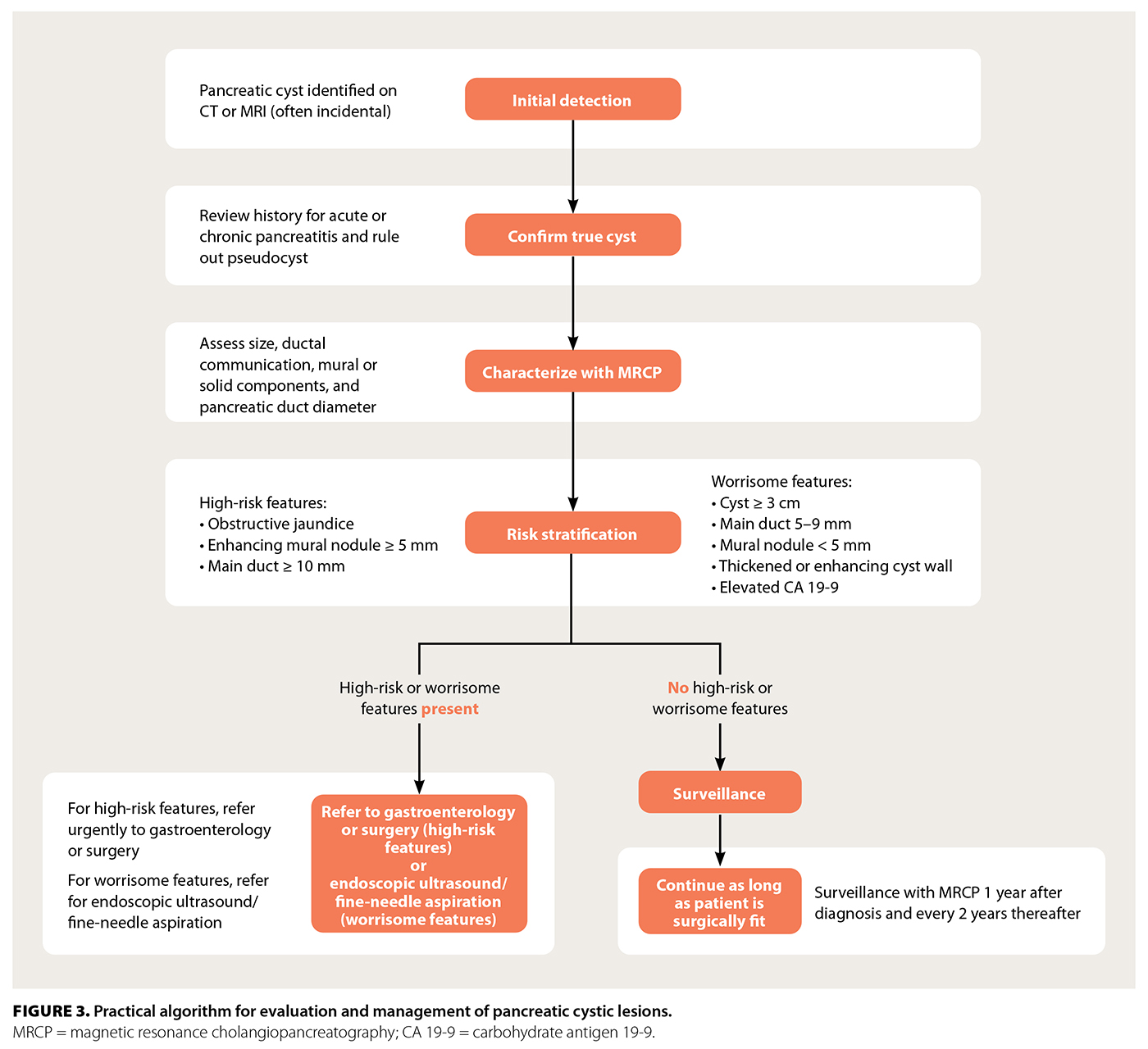 Management of pancreatic cystic lesions is guided by the type of cyst, malignant potential, and presence of high-risk features, with the overarching goal of identifying lesions at risk for invasive carcinoma while avoiding unnecessary pancreatic surgery. In general, pancreatic cystic lesions with a known high risk of malignancy, such as main-duct or mixed-type IPMNs, mucinous cystic neoplasms, and solid pseudopapillary neoplasms, should be referred for surgical resection if the patient is an acceptable surgical candidate.[3,5,14,22] Surgery is also indicated when a cyst demonstrates high-risk imaging or clinical features, including main pancreatic duct dilation 10 mm or larger, an enhancing mural nodule 5 mm or larger, obstructive jaundice related to the cyst, or cytology showing high-grade dysplasia or malignancy.[2-4,23] In addition, symptomatic cysts causing pancreatitis, gastric outlet obstruction, or persistent pain warrant resection regardless of malignant potential.[2,5] A practical stepwise approach to management is outlined in Figure 3.
For asymptomatic lesions without high-risk features, active surveillance is preferred, because the risk of progression to advanced neoplasia remains low. MRCP is the recommended modality for longitudinal follow-up.[2-4] The frequency and duration of surveillance vary across major guidelines and depend primarily on cyst size, morphology, and interval stability; these recommendations are summarized in the Table. In general, MRCP is conducted 1 year after diagnosis and then every 2 years thereafter for those without worrisome features. Surveillance may be discontinued if the patient becomes unfit or unwilling to undergo pancreatic surgery.[2-5,23]
Patients who have undergone partial pancreatic resection for IPMN require ongoing follow-up of the pancreatic remnant, because metachronous cysts can develop even years later. Annual MRCP or endoscopic ultrasonography is generally recommended, particularly when the resected IPMN harbored high-grade dysplasia, because these individuals remain at elevated risk of subsequent pancreatic cancer.[24,25]
Overall, the prognosis for noninvasive cystic lesions is excellent when managed according to evidence-based pathways. Importantly, studies have shown that pancreatic cancers detected during structured surveillance are more often found at early, surgically curable stages, which reinforces the value of guideline-directed monitoring in appropriately selected patients.[25]
Key points: Management
- Lesions with high malignant potential (main-duct/mixed-type intraductal papillary mucinous neoplasm (IPMN), mucinous cystic neoplasm, solid pseudo-papillary neoplasm) or high-risk imaging/clinical features should be referred for surgical resection in appropriate surgical candidates.
- Asymptomatic lesions without high-risk features are managed with active surveillance, typically magnetic resonance cholangiopancreatography, at 1 year after diagnosis and every 2 years thereafter [Figure 3].
- After partial pancreatectomy for IPMN, lifelong annual surveillance of the pancreatic remnant is required, given the risk of metachronous disease, especially if high-grade dysplasia was present.
Summary
Pancreatic cystic lesions are increasingly detected with modern imaging, and most are benign or indolent. However, a subset, particularly mucinous and main-duct lesions, carry a measurable risk of malignant transformation. MRCP is the preferred imaging modality for characterization and surveillance, while endoscopic ultrasonography with cyst-fluid analysis provides additional diagnostic precision when indicated. The presence of high-risk features such as mural nodules, main duct dilation, or obstructive jaundice should prompt referral to a gastroenterologist or pancreatic centre for surgical evaluation. For most asymptomatic cysts that lack these features, structured surveillance following established guideline intervals is safe and effective. Early recognition, risk stratification, and coordinated follow-up between primary care and specialist teams are key to ensuring timely detection of malignant change while minimizing unnecessary intervention.
Competing interests
None declared.
This article has been peer reviewed.

This work is licensed under a Creative Commons Attribution-NonCommercial-NoDerivatives 4.0 International License.
References
1. Laffan TA, Horton KM, Klein AP, et al. Prevalence of unsuspected pancreatic cysts on MDCT. Am J Roentgenol 2008;191:802-807. https://doi.org/10.2214/AJR.07.3340.
2. Elta GH, Enestvedt BK, Sauer BG, Lennon AM. ACG clinical guideline: Diagnosis and management of pancreatic cysts. Am J Gastroenterol 2018;113:464-479. https://doi.org/10.1038/ajg.2018.14.
3. Ohtsuka T, Fernández-del Castillo C, Furukawa T, et al. International evidence-based Kyoto guidelines for the management of intraductal papillary mucinous neoplasm of the pancreas. Pancreatology 2024;24:255-270. https://doi.org/10.1016/j.pan.2023.12.009.
4. Megibow AJ, Baker ME, Morgan DE, et al. Management of incidental pancreatic cysts: A white paper of the ACR Incidental Findings Committee. J Am Coll Radiol 2017;14:911-923. https://doi.org/10.1016/j.jacr.2017.03.010.
5. European Study Group on Cystic Tumours of the Pancreas. European evidence-based guidelines on pancreatic cystic neoplasms. Gut 2018;67:789-804. https://doi.org/10.1136/gutjnl-2018-316027.
6. Fung CI, Bigam DL, Wong CKW, et al. Recommendations for the management of incidental pancreatic findings in adults by the Canadian Association of Radiologists Incidental Findings Working Group. Can Assoc Radiol J 2022;73:312-319. https://doi.org/10.1177/08465371211021079.
7. Feldman M, Friedman LS, Brandt LJ. Sleisenger and Fordtran’s gastrointestinal and liver disease e-book: Pathophysiology, diagnosis, and management. 11th ed. Philadelphia, PA: Elsevier; 2020.
8. Bergin D, Ho LM, Jowell PS, et al. Simple pancreatic cysts: CT and endosonographic appearances. Am J Roentgenol 2002;178:837-840. https://doi.org/10.2214/ajr.178.4.1780837.
9. Adsay NV, Hasteh F, Cheng JD, et al. Lymphoepithelial cysts of the pancreas: A report of 12 cases and a review of the literature. Mod Pathol 2002;15:492-501. https://doi.org/10.1038/modpathol.3880553.
10. Nasr J, Sanders M, Fasanella K, et al. Lymphoepithelial cysts of the pancreas: An EUS case series. Gastrointest Endosc 2008;68:170-173. https://doi.org/10.1016/j.gie.2008.02.044.
11. Assifi MM, Nguyen PD, Agrawal N, et al. Non-neoplastic epithelial cysts of the pancreas: A rare, benign entity. J Gastrointest Surg 2014;18:523-531. https://doi.org/10.1007/s11605-014-2459-7.
12. Yun W-G, Seo Y, Han Y, et al. Reappraisal of surgical indication of mucinous cystic neoplasm of the pancreas: A systematic review and meta-analysis. Int J Surg 2025. https://doi.org/10.1097/JS9.0000000000004031.
13. Lee LS, Gardner TB. Pancreatic cysts and pancreatitis in the older adult. Am J Gastroenterol 2025;120(Suppl 10):S27-S33. https://doi.org/10.14309/ajg.0000000000003641.04.
14. Law JK, Ahmed A, Singh VK, et al. A systematic review of solid-pseudopapillary neoplasms: Are these rare lesions? Pancreas 2014;43:331-337. https://doi.org/10.1097/MPA.0000000000000061.
15. Stark A, Donahue TR, Reber HA, Hines OJ. Pancreatic cyst disease: A review. JAMA 2016;315:1882-1893. https://doi.org/10.1001/jama.2016.4690.
16. Smith ZL, Satyavada S, Simons-Linares R, et al. Intracystic glucose and carcinoembryonic antigen in differentiating histologically confirmed pancreatic mucinous neoplastic cysts. Am J Gastroenterol 2022;117:478-485. https://doi.org/10.14309/ajg.0000000000001623.
17. Raman A, Lennon AM. Cyst fluid biomarkers – Diagnosis and prediction of malignancy for cystic lesions of the pancreas. Visc Med 2018;34:178-181. https://doi.org/10.1159/000490137.
18. Del Chiaro M, Segersvärd R, Pozzi Mucelli R, et al. Comparison of preoperative conference-based diagnosis with histology of cystic tumors of the pancreas. Ann Surg Oncol 2014;21:1539-1544. https://doi.org/10.1245/s10434-013-3465-9.
19. Das KK. The “next generation” of pancreatic cyst fluid biomarkers? Gastroenterology 2023;164:21-23. https://doi.org/10.1053/j.gastro.2022.10.023.
20. Maguchi H, Tanno S, Mizuno N, et al. Natural history of branch duct intraductal papillary mucinous neoplasms of the pancreas: A multicenter study in Japan. Pancreas 2011;40:364-370. https://doi.org/10.1097/MPA.0b013e31820a5975.
21. Tanno S, Nakano Y, Koizumi K, et al. Pancreatic ductal adenocarcinomas in long-term follow-up patients with branch duct intraductal papillary mucinous neoplasms. Pancreas 2010;39:36-40. https://doi.org/10.1097/MPA.0b013e3181b91cd0.
22. Nilsson LN, Keane MG, Shamali A, et al. Nature and management of pancreatic mucinous cystic neoplasm (MCN): A systematic review of the literature. Pancreatology 2016;16:1028-1036. https://doi.org/10.1016/j.pan.2016.09.011.
23. Vege SS, Ziring B, Jain R, et al. American Gastroenterological Association Institute guideline on the diagnosis and management of asymptomatic neoplastic pancreatic cysts. Gastroenterology 2015;148:819-822. https://doi.org/10.1053/j.gastro.2015.01.015.
24. Majumder S, Philip NA, Singh Nagpal SJ, et al. High-grade dysplasia in resected main-duct intraductal papillary mucinous neoplasm (MD-IPMN) is associated with an increased risk of subsequent pancreatic cancer. Am J Gastroenterol 2019;114:524-529. https://doi.org/10.1038/s41395-018-0403-2.
25. de la Fuente J, Lui J, Lennon RJ, et al. Pancreatic cancer is more frequently early stage at diagnosis in surgically resected intraductal papillary mucinous neoplasms with preoperative surveillance. Gastro Hep Adv 2022;1:1099-1107. https://doi.org/10.1016/j.gastha.2022.07.004
Dr Kakkar is an internal medicine resident in the Faculty of Medicine at the University of British Columbia. Dr Sugrue is a clinical assistant professor in the Faculty of Medicine at UBC and the Department of Radiology at Vancouver General Hospital. Dr Gan is a clinical associate professor in the Department of Medicine at UBC and the Division of Gastroenterology at Vancouver General Hospital.
Corresponding author: Dr S. Ian Gan, ian.gan@vch.ca.