Diabetic ketoacidosis in children and adolescents: An update and revised treatment protocol
Issue: BCMJ,
vol. 52 , No. 1 , January February 2010 ,
Pages 24-31 Clinical Articles
ABSTRACT: British Columbia has an estimated 150 to 200 new cases of type 1 diabetes in children annually. In these cases, 10% to 20% of patients will present in diabetic ketoacidosis (DKA). DKA is associated with significant fluid and biochemical derangements, necessitating a thoughtful, structured approach to its management. Recent gains have been made in knowledge about the pathophysiology and medical care of DKA and its most significant complication, cerebral edema. In response, BC Children’s Hospital has devised an updated medical protocol for managing DKA in infants, children, and adolescents that conforms to new international consensus guidelines. The protocol assists the medical practitioner in calculating fluid and electrolyte replacement needs for individual patients and outlines a plan for initial assessment and ongoing monitoring. Accompanying resources have also been developed to aid nursing, laboratory, and pharmacy colleagues to ensure that all children presenting with DKA in this province are managed following scientifically established guidelines.
Standardized pediatric-specific treatment is required to ensure safe correction of metabolic derangements associated with DKA.
BCMJ.org Health Notes patient information sheet is available for this article.
Canada has one of the highest rates of type 1 diabetes (T1D) in the world. The estimated incidence of T1D in Canadian children aged 0 to 14 years is 21.7 per 100000 per year.[1] Using 2008 census data,[2] prevalence in this age group in British Columbia is estimated to be about 1029 established cases of T1D or about 150 new cases per year. Much publicity has been given to the rising incidence of type 2 diabetes (T2D) in youth and young adults in North America, a phenomenon that we are also observing in our province, but the fact that there has also been a 2% to 3% annual increase in the incidence of T1D over the past two decades is not as well publicized.[3]
Diagnosis of diabetes and DKA in children
The diagnosis of T1D is generally straightforward in the pediatric patient. The Canadian Diabetes Association’s 2008 Clinical Practice Guidelines[4] state that a random blood glucose greater than or equal to 11.1 mmol/L in the face of typical symptoms (polyuria, polydipsia, weight loss, and fatigue) is sufficient to make the diagnosis of diabetes.
The guidelines further emphasize that the diagnosis of T1D in children should be confirmed at presentation and not be delayed by waiting for the results of fasting blood glucose levels; an oral glucose tolerance is rarely indicated in this setting.
Despite awareness campaigns targeting both lay and professional audiences, an estimated 10% to 20% of children with new-onset T1D still present in diabetic ketoacidosis (DKA). As well, children with established T1D can develop recurrent DKA because of deliberate or accidental insulin omission. It should also be noted that children and youth with T2D can present with DKA, and that the presence of ketones does not invariably point to the diagnosis of T1D.
The criteria for the diagnosis of DKA are relatively clear-cut:
• Hyperglycemia: blood glucose =11.1 mmol/L.
• Acidosis: venous pH <7.3 and/or bicarbonate <15 mmol/L.
• Ketosis: presence of ketones in the blood, urine, or both.
DKA cases can be roughly divided further into mild (pH 7.20–7.29, bicarbonate 10–14), moderate (pH 7.10–7.29, bicarbonate 5–9), and severe (pH <7.10, bicarbonate <5). Endocrinologists may feel comfortable treating mild DKA in reliable patients with known T1D at home or as outpatients.
The majority of children with moderate DKA and all children with severe DKA should be treated in a medical facility, optimally by a pediatric endocrinologist, pediatrician, or other practitioner familiar with the unique issues that arise in DKA in the young.
Clinically, children in DKA present with the following: dehydration of up to 10% of their body weight; Kussmaul respirations; nausea, vomiting, and abdominal pain; and a progressive decrease in consciousness.
Cerebral edema: Pathophysiology and risk factors
The major mortality factor associated with DKA in infants, children, and adolescents is cerebral edema (DKA-CE). This phenomenon has been documented on cranial CT scans prior to initiation of treatment, and it has been observed in adults as well. Approximately 0.5% to 1.5% of children presenting with DKA will develop clinically evident DKA-CE.[5]
In a recent Canadian surveillance study, 23% of children developing DKA-CE died, and another 15% survived with neurological complications.[6] DKA-CE accounts for 70% to 80% of diabetes-related deaths in children under 12 years. Other causes of death include electrolyte disturbances, shock, cerebral venous thrombosis, and pulmonary edema.
The exact pathophysiology underlying DKA-CE remains unclear.[7] The accumulation of water in the brain may be due to the presence of so-called idiogenic osmoles, small organic compounds that are formed in the intracellular space as a defensive response to increasing osmolality in the extracellular compartment.
When fluid resuscitation is initiated, the extracellular space becomes relatively hypotonic compared with the intracellular space, resulting in an influx of water into neurons. Activation of the sodium-hydrogen exchanger mechanism, perhaps by insulin, vasopressin, or both may also lead to an influx of sodium ions (followed by water) into brain cells.
Finally, it is hypothesized that DKA-CE may be due to a vasogenic mechanism representing reperfusion of hypoperfused tissue. Taken together, these factors would suggest that a thoughtful approach is necessary in the administration of fluids, electrolytes, and insulin.
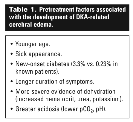
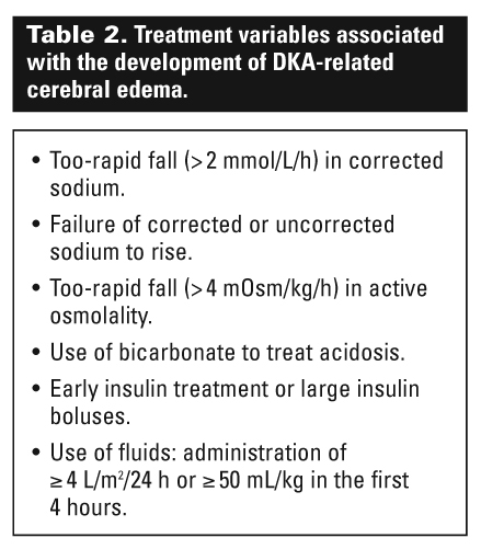
A number of pretreatment (Table 1) and treatment-associated (Table 2) risk factors for the development of DKA-CE have been described.[8] In general, younger children presenting with new-onset diabetes who have had symptoms of long duration and increased severity, as well as more pronounced dehydration and acidosis, are at highest risk of developing DKA-CE.
Alternatively, sicker-appearing children may be more likely to elicit anxiety in the medical team, leading to hasty treatment decisions. Treatment regimens associated with over-zealous administration of fluids have likewise been implicated in the development of DKA-CE in a number of studies.[8]
Most recently, a case-control study in children from the UK demonstrated for the first time that administration of insulin within the first hour of treatment was independently associated with up to a 12.7-fold risk of developing DKA-CE.[9]
Most episodes of DKA-CE occur 4 to 12 hours after treatment has started. Timely recognition of the signs and symptoms leading to prompt initiation of treatment of DKA-CE is imperative. Most children will complain of headache and begin vomiting prior to developing an altered state of consciousness, decreased response to pain, decorticate or decerebrate posturing, cranial nerve palsies, and Cushing’s triad (hypertension, bradycardia, and abnormal respiratory pattern).
Emergency treatment includes elevation of the head of the bed, a decrease in fluid rate by one-third, and administration of mannitol (0.5–1 g/kg over 20 minutes) or 3% sodium chloride (5–10 mL/kg IV over 30 minutes). Intubation with mild hyperventilation to keep the pCO2 above 22 mm Hg may also be required.
Protocols for treating DKA
Because of the high rates of mortality and morbidity associated with DKA in children, young patients should be managed using standardized pediatric-specific treatment protocols to ensure safe correction of metabolic derangements while minimizing the risk of development of DKA-CE.[4]
The underlying principle of modern protocols is to provide even rehydration and restoration of body fluid and electrolyte deficits over a 48-hour period. This requires a meticulous approach to calculating intravenous fluid composition and rates of administration.
In 1996, the Endocrinology and Diabetes Unit at BC Children’s Hospital (BCCH) developed a DKA protocol based on the international guidelines and evidence-based knowledge that were available at that time. A companion article published in the BCMJ detailed the 1996 protocol and its rationale.[10]
Since then, new information has been published about the pathophysiology of DKA and the complications of its treatment, including DKA-CE. This has necessitated the updating of DKA protocols by a number of pediatric endocrine societies and academic institutions.
In 2007, the International Society for Pediatric and Adolescent Diabetes (ISPAD) published its Clinical Practice Consensus Guidelines for DKA (updated again in 2009).[8] The ISPAD guidelines are considered the current gold standard internationally, and the recommendations published therein have been endorsed by most major subspecialist societies.
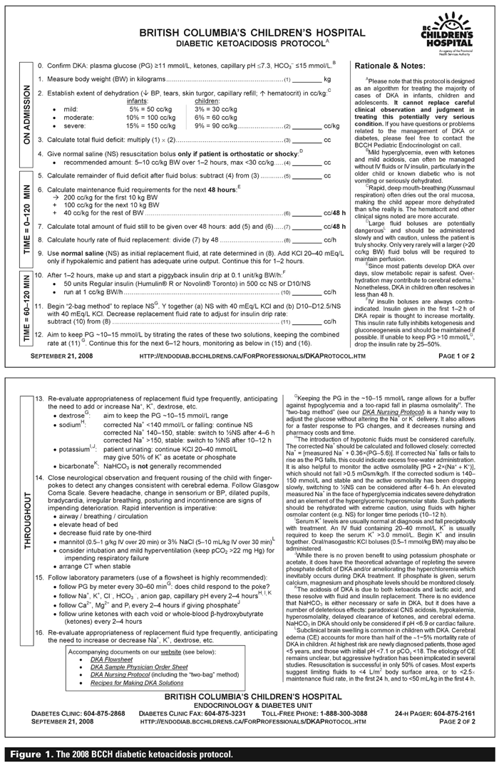
In late 2008, the staff of the BCCH Endocrinology and Diabetes Unit published a revision of their DKA protocol to bring it into conformity with the ISPAD guidelines. The protocol is now available online[11] as part of the BCCH DKA Toolkit, which includes the following:
• A medical protocol for DKA (see Figure 1).
• A nursing protocol for DKA.
• A flowsheet for managing DKA (see Figure 2).
• A sample physician order sheet for DKA.
• Recipes for making DKA solutions.
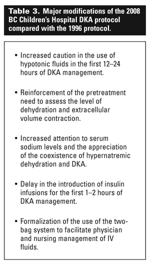
The five major modifications of the 1996 protocol are summarized in Table 3. As in the original protocol, the revised protocol divides the management of DKA into three periods, based on the need for monitoring and the composition of fluids to be administered:
• On admission: attend to ABCs (airway, breathing, and circulation); complete clinical assessment; provide fluid bolus if necessary.
• The first 1 to 2 hours: start normal saline to begin rehydration.
• Thereafter: begin insulin infusion; add potassium and eventually dextrose to IV fluids; continue clinical and biochemical monitoring and adjustments.
The nursing protocol has been designed to provide nursing personnel with a more detailed explanation of how to perform initial and follow-up assessments of the DKA patient, how to prepare and manage the IV fluids and insulin infusion, and how to coordinate the necessary laboratory and bedside blood testing.
Should nursing or pharmacy staff require assistance in making nonstandard IV solutions, the BCCH DKA Toolkit has a recipes page that explains how to prepare these using commercially available products.
In the emergency room and after
As with all sick patients, attending to the ABCs is the first priority. It is important to document the initial Glasgow Coma Scale score for use as a baseline. Since all fluid calculations in the protocol are based on the patient’s current weight, it is imperative that an accurate weight be obtained on all patients.
An estimation of dehydration should be made, based on clinical criteria (tears, skin turgor, or capillary refill) and laboratory results (hematocrit, urea, potassium). Note that urine output cannot be relied upon as an accurate measure of dehydration because of the obligate diuresis of hyperglycemia.
Similarly, with the mouth-breathing of Kussmaul respirations, oral mucous membranes will often appear drier than the patient’s actual level of fluid deficit. A normal blood pressure is reassuring, but nearly all children in the ER will be anxious and tachycardic.
It has become apparent that the biochemical picture of children presenting in DKA depends to a substantial degree on the type of fluid (milk, water, pop, fruit juice) and amount (if any) the child has been receiving in the time before presentation, so it is important to ask about this.
Careful history-taking is also essential to help identify any events that might have led to recurrent DKA in a patient with known T1D. Finally, the physician must remember that underlying infections or illness can trigger or accelerate the development of DKA.
Once a large-bore intravenous cannula has been placed and laboratory samples have been obtained to provide the baseline values needed (glucose, electrolytes, capillary blood gas, urea, creatinine, CBC, A1c, blood or urine ketones), fluid replacement can begin.
If the patient shows evidence of cardiovascular instability, such as severe tachycardia, hypotension, or decreased capillary refill, a fluid bolus of normal saline (5–10 cc/kg) can be given over 30 to 60 minutes; very rarely, the shocky patient may require a second or third fluid push.
The BCCH DKA protocol has a formula for calculating fluid rates, depending on the patient’s weight and level of dehydration. In the initial 1 to 2 hours, normal saline is given. Thereafter, assuming the patient has been documented to have urine output, potassium (as chloride) should be added to the rehydration fluid. Insulin is started after the initial 1 to 2 hours at a dosage of 0.1 U/kg/h; instructions for making this are in the medical and nursing protocols.
Once started, it is important that the insulin not be interrupted. IV insulin has a very short circulating half-life, and the metabolic derangements of DKA, which are normally suppressed by the insulin infusion, can restart within minutes of its discontinuation. If the insulin causes the patient to become hypoglycemic, it is preferable to add dextrose to the IV solutions using the two-bag system. Boluses of insulin should be avoided.
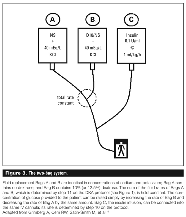
The two-bag system
Once the patient is receiving fluids and then insulin, the blood glucose will fall, often quite rapidly. The goal is to maintain the blood glucose in the 10 to 15 mmol/L range over the first day or so, to provide a buffer against the development of hypoglycemia. For this reason, dextrose should be added to the replacement fluids at this point.
To minimize turnaround time for fluid adjustments, our institution introduced the two-bag system a number of years ago, based on results published by a large pediatric centre.[12] In short, two bags of intravenous fluids, identical in their electrolyte composition and differing only in their dextrose concentration, are run in parallel through the same cannula (see Figure 3).
The total fluid rate from these two bags—determined by the protocol—will be constant, and the final concentration of dextrose can be altered simply by juggling the rates of the two bags. The insulin infusion from a third bag can be connected into the same cannula.
The two-bag system is easy to institute, uses commercially available solutions, and has been shown to decrease the time needed to make a change in IV rates, to decrease the number of IV bags used during an admission, and to decrease the cost of IV solutions used.[12]
Monitoring the DKA patient
The patient in DKA will require frequent biochemical monitoring to ensure a smooth, safe correction of the metabolic derangements. The use of a flowsheet is indispensable in tracking the progress of a patient. Following the anion gap and capillary pH allows for monitoring of the correction of the acidosis.
Special attention should be paid to the rise or fall of the sodium level as corrected for glucose. This is calculated as follows: corrected Na+ = [measured Na+ + 0.36(glucose–5.6)]. A low corrected sodium that is not rising, or a corrected sodium that is dropping rapidly (>2 mmol/L/h), suggests excess administration of free water.
Some experts have proposed following the active osmolality, a measure of the osmotically active molecules (which does not include urea) in DKA as follows: active osmolality = [2(Na+ + K+) + glucose]. A rapid fall in the active osmolality (>4 mOsm/kg/h) has also been associated with the development of DKA-CE.
Traditionally, ketones have been assessed by using semiquantitative urine dipsticks (measuring acetoacetate, which is actually present to a lesser degree in DKA). In recent years, quantitative blood beta-hydroxybutyrate levels have been used increasingly to follow the development and resolution of ketosis.
Blood beta-hydroxybutyrate rises more rapidly and corrects more quickly than urine ketones and is thus a more sensitive indicator of impending DKA. Many patients using insulin pumps are trained to check themselves using a home monitor to detect early ketones associated with infusion-site problems. A blood beta-hydroxybutyrate greater than or equal to 0.4 mmol/L is abnormal in children with diabetes. Table 4 provides a comparison of urine and blood ketones.[13]
Caveats
While it is generally straightforward to diagnose and treat DKA, caution must be taken when treating a child in DKA to ensure that comorbidities are not missed. Many children presenting in DKA have abdominal pain, tachypnea, and polyuria; it is important to ascertain with time whether these are part of the DKA picture or there is an underlying appendicitis, pneumonia, or urinary tract infection.
Fever is not seen in uncomplicated DKA and should always be investigated. However, white blood cell counts are often quite elevated in DKA with a profound left shift, due to elevated plasma catecholamines; this normally resolves within the first 4 to 6 hours of treatment.
It has been recognized increasingly that many children presenting with DKA-like symptoms may have acoexistent—and sometimes predominant—element of hypernatremic dehydration, the hyperglycemic hyperosmolar state, or both. This is observed especially in children with T2D, as well as those with T1D who have consumed large amounts of salt- or carbohydrate-containing fluids prior to presentation.
In these children, hypernatremia (corrected Na+ >150 mmol/L), hyperglycemia (>33 mmol/L), and/or hyperosmolality (>320 mOsm/kg) may be present, but the acidosis is mild (pH >7.30, bicarbonate > 15 mmol/L), and ketones are absent or only mildly elevated (blood beta-hydroxybutyrate <1 mmol/L). In such cases, treatment must be modified to address the unique biochemical disturbance of the patient.
The ketoacidosis of DKA resolves with fluid and insulin treatment. Bicarbonate should not be used in children with DKA, except in the very rare situation where profound acidosis is causing decreased cardiac output. The use of bicarbonate has been associated with an increased risk of DKA-CE, paradoxical CNS acidosis, hyperosmolality, delayed correction of acidosis, and longer hospital stays.[8]
As well, the use of phosphate-containing solutions in DKA remains controversial. Prospective studies have not identified a benefit with the use of phosphate, but most experts would consider substituting some of the potassium chloride in the replacement fluids with potassium phosphate in the face of weakness or severe hypophosphatemia.[8] In this case, the patient must be monitored for the development of hypocalcemia.
Occasionally, patients treated according to the DKA protocol do not exhibit an increase in pH after the first 4 to 6 hours of treatment. Almost invariably, this is due to an error in the preparation of the insulin infusion. In this case, a new insulin bag should be made.
Existing DKA protocols, including the BCCH protocol, provide an algorithm for treating the majority of cases of DKA in infants, children, and adolescents based on our best current understanding of research and the medical literature. However, no protocol has been designed that completely eliminates the occurrence of DKA-CE, and no protocol can replace careful clinical observation and judgment when treating this potentially very serious condition.
Recurrent DKA
Most pediatric endocrinologists will maintain that the vast majority of recurrent DKA episodes are preventable. The physician should bear in mind that recurrent DKA is the result of insulin omission, either deliberate or accidental, until proven otherwise.
An A1c taken at admission, and perhaps an insulin level, will help identify the patient with recurrent bouts of “gastroenteritis” who is in reality in borderline metabolic control or who has been missing insulin injections (or both). Insulin omission is frequently associated with eating disorders in teenage girls, depression in children or youth, insufficient parental supervision, or a poor psychosocial situation. In these instances, it is often beneficial to solicit the help of a social worker, counselor, or psychiatrist before discharge.
In some instances, parents or patients will mistakenly discontinue all insulin on sick days because of a fear of hypoglycemia with poor oral intake. This situation may also lead to hyperglycemia and DKA. Family education around proper sick-day management, with reinforcement at subsequent visits, should help to prevent this problem.
With the rise in use of insulin pumps in the pediatric population (approximately 30% to 40% of children who have diabetes in BC are on an insulin pump), a new cause of DKA is being observed: insulin pump infusion-site problems (and, much more uncommonly, insulin pump malfunction).
Since pumps carry only rapid-acting insulin analogs, any disruption in insulin delivery can lead to rapid development (within 2 to 4 hours) of hyperglycemia and ketosis. Home monitoring of ketones, rapid replacement of dislodged infusion sites, and the administration of a correction dose of rapid-acting insulin (preferably by pen or syringe if ketones are present) are essential.
Family members should be educated about managing infusion-site problems at the time of pump initiation, with reinforcement of pump training at subsequent visits.
Coming off protocol
While the protocol is designed to correct DKA over a 48-hour period, many children are metabolically corrected more quickly. Once the acidosis has resolved (pH >7.3 and anion gap normal) and the patient is feeling well and is ready to begin eating and drinking, the use of subcutaneous insulin can be started or re-established. This is most easily done at breakfast or dinner.
The patient will require a dose of intermediate-acting (e.g., NPH) or basal (e.g., glargine or detemir) insulin, as well as a dose of short-acting (e.g., regular) or rapid-acting (e.g., lispro, aspart, or glulisine) preprandial insulin. The insulin drip should be discontinued 15 to 30 minutes after the first injection of rapid-acting insulin or 60 to 120 minutes after regular insulin. The physician may choose to continue the IV fluids (without glucose) in the patient who still has a mild fluid deficit.
Summary
The management of pediatric DKA in our province often begins in local emergency rooms with primary care providers who see this condition only rarely. Yet it is the medical care that children receive in the first hours that can have the greatest impact on their outcome and survival.
It is essential for all emergency rooms and associated medical personnel to have a plan in place for dealing with this relatively uncommon condition, including access to the necessary medical supplies and diagnostic equipment needed to make a rapid and accurate diagnosis.
A thoughtful plan of action must be formulated to maximize patient safety. It must be recognized that DKA is treated very differently in children and adults, particularly with respect to fluid administration. Pediatric tertiary care centres such as BCCH stand ready to assist local and regional hospitals and medical staff in dealing with pediatric DKA at all times.
Ultimately, the goal is to decrease the incidence of DKA in children by educating the public about the signs and symptoms of diabetes, reminding T1D families about avoidance of recurrent DKA, and training professionals to make an earlier diagnosis of diabetes in children presenting with suspicious symptoms.
Acknowledgments
The BCCH DKA Protocol Toolkit was revised by the pediatric endocrinologists from the Endocrinology and Diabetes Unit at BC Children’s Hospital, who are grateful for the input received from colleagues in Nursing, Intensive Care, General Pediatrics, Emergency Medicine, Laboratory Medicine, and Pharmacy.
Competing interests
None declared.
References
1. International Diabetes Federation. IDF diabetes atlas. 3rd ed. 2008. www.eatlas.idf.org (accessed 23 October 2009).
2. Statistics Canada. Population by sex and age group, by province and territory, 2008. www40.statcan.gc.ca/l01/cst01/demo31a-eng.htm (accessed 23 October 2009).
3. Gale EA. The rise of childhood type 1 diabetes in the 20th century. Diabetes 2002;51:3353-3361.
4. Canadian Diabetes Association. 2008 Clinical practice guidelines for the prevention and management of diabetes in Canada. Can J Diabetes 2008;32(suppl. 1). www.diabetes.ca/for-professionals/resources/2008-cpg/ (accessed 23 October 2009).
5. Dunger DB, Sperling MA, Acerini CL, et al. European Society for Paediatric Endocrinology/Lawson Wilkins Pediatric Endocrinology Society consensus statement on diabetic ketoacidosis in children and adolescents. Pediatrics 2004;113:133-140.
6. Lawrence SE, Cummings EA, Gaboury I, et al. Population-based study of incidence and risk factors for cerebral edema in pediatric diabetic ketoacidosis. J Pediatr 2005;146:688-692.
7. Glaser N, Barnett P, McCaslin I, et al. Risk factors for cerebral edema in children with diabetic ketoacidosis. N Engl J Med 2001;344:264-269.
8. Wolfsdorf J, Craig ME, Daneman D, et al; International Society for Pediatric and Adolescent Diabetes. ISPAD clinical practice consensus guidelines 2009 compendium: Diabetic ketoacidosis. Pediatr Diabetes 2009;10(suppl 12):118-133. www.ispad.org/FileCenter.html/CategoryID=5 (accessed 23 October 2009).
9. Edge JA, Jakes RW, Roy Y, et al. The UK case-control study of cerebral oedema complicating diabetic ketoacidosis in children. Diabetologia 2006;49:2002-2009.
10. Metzger DL. A provincial protocol for the treatment of diabetic ketoacidosis in children. BCMJ 1998;40:110-115.
11. BC Children’s Hospital Endocrinology and Diabetes Unit. Diabetic ketoacidosis protocol toolkit. http://endodiab.bcchildrens.ca/ForProfessionals/DKAProtocol.htm (accessed 23 October 2009).
12. Grimberg A, Cerri RW, Satin-Smith M, et al. The “two-bag” system for variable intravenous dextrose and fluid administration: Benefits in diabetic ketoacidosis management. J Pediatr 1999;134:376-378.
13. Brink S, Laffel L, Likitmaskul S, et al.; International Society for Pediatric and Adolescent Diabetes. ISPAD clinical practice consensus guidelines 2009 compendium: Sick day management in children and adolescents with diabetes. Pediatr Diabetes 2009;10(suppl 12):146-153. www.ispad.org/FileCenter.html?CategoryID=5 (accessed 23 October 2009).
Dr Metzger is a staff endocrinologist in the Endocrinology & Diabetes Unit at British Columbia’s Children’s Hospital and a clinical professor in the Department of Pediatrics at the University of British Columbia.
