CT colonography: A new technique for colorectal cancer screening
Issue: BCMJ,
vol. 50 , No. 4 , May 2008 ,
Pages 206-211 Clinical Articles
Although optical colonoscopy is the criterion standard for colorectal cancer screening, CT colonography is now accepted to be useful when screening average-risk patients. Earlier studies that indicated poor sensitivity for CT colonography have now been largely refuted. Recent improvements in workstation design and advances that permit primary 3D viewing of images have increased screening accuracy. Access to CT colonography is still somewhat limited, but some advantages of CT colonography over optical colonoscopy include the lack of side effects from sedation and low perforation rates. Disadvantages of screening with CT colonography include the need to schedule a subsequent optical colonoscopy if images indicate a biopsy or polypectomy is required, and the reduced ability of CT colonography to identify small polyps which rarely, but occasionally, are significant.
Average-risk patients may be candidates for a less invasive imaging technology.
Colorectal cancer (CRC) is the third most common malignancy in Canada and the number one cause of cancer death among nonsmokers in North America.[1] The incidence increases significantly after age 50.
More than 700 people die of CRC annually in BC alone, each losing an estimated 10 years of productive life, for a total yearly provincial loss of over 7000 productive years.[2] Three-quarters of all CRC occurs in asymptomatic average-risk individuals, that is, patients older than 50, without a family history of the disease, and with no other risk factors such as inflammatory bowel disease or familial polyposis.
CRC: The ideal disease for screening
Because CRC exists initially as premalignant polyps in virtually all cases, and because early CRC is more curable than late CRC (90% cure rates when patient is asymptomatic vs 50% cure rates when patient is symptomatic), this disease is ideally suited for a screening program. However, two Canadian studies have shown very poor compliance rates for CRC screening.
A study by Rabeneck and Paszat[3] of nearly 1 million screen-eligible Ontario patients age 50 to 59 determined that fewer than 20.5% had undergone any form of CRC screening during a 6-year follow-up period.
A more recent phone survey[4] of 1808 Alberta men and women between age 50 and 74 determined that only 13.2% of these average-risk patients had been advised by a physician to undergo CRC screening and only 3% had undergone endoscopy within the past 5 years, despite the fact that screening for CRC is supported by both the Canadian Cancer Society and the American Cancer Society.
Multiple procedures for CRC screening are available, including digital rectal examination, fecal occult blood testing, double-contrast barium enema, and flexible sigmoidoscopy. Other procedures on the horizon include video colon capsules and DNA testing.
However, at present optical colonoscopy (OC) remains the criterion standard and should be used when assessing any new techniques. One such technique, CT colonography (CTC), is now accepted to have an important role to play in screening average-risk patients.
At present, access to CTC is still somewhat limited, although there are now a number of public imaging facilities providing the service in BC. Any local population-based impact on CRC screening, however, is likely to be some years away.
CT Colonography
CT colonography was first introduced by Vining and Gelfand in 1994. CTC is now the preferred name, although “virtual colonoscopy” is also commonly used. Since its introduction, CTC has benefited immensely from technological improvements such as multidetector CT scanners and improved workstations.
At present an optimal CTC examination still requires a bowel cleansing regimen identical to that used for colonoscopy, with the addition of dilute barium and a cholecystographic agent (e.g., Telepaque) taken orally. Barium, by becoming incorporated into any remaining stool, helps differentiate polyps from stool.
Telepaque, by increasing the density of any residual colonic fluid, helps reveal polyps that otherwise may remain isodense within fluid and not be visualized. Other means of “tagging” stool in concert with advanced image processing techniques may mean that in future less or no colonic preparation will be required.[5]
The CTC procedure begins when a rectal tube is inserted and the colon is insufflated manually with carbon dioxide, which is preferred to room air because it has a much greater rate of resorption, and thus diminishes patient discomfort.[6]
Optimal colon preparation and distention is absolutely vital to ensure an adequate study. Inadequate prep and colonic distention are the most common causes of false-negative examinations.[7]
The patient is scanned in both the supine and prone positions. In problem cases, the decubitus position may be used as well. These maneuvers improve the probability of adequately distending all colonic segments, shift any residual fluid from segment to segment (unmasking polyps that may be hidden within a fluid column), and help to differentiate stool, which tends to be mobile, from polyps, which remain static.
Intravenous contrast is not generally used for CTC examinations as it is not felt that the increased but still very small risk associated with possible contrast reactions is justified in the well-prepped colon. Spasmolytics are also not routinely utilized as several studies have shown them to be of little value.[8]
A Toshiba 64 multislice scanner is used by the authors. Scans are acquired at 0.5 mm resolution and reconstructed at 2 mm collimation at 25 to 30 mAs and 120 kVp. Such thin slices are required to diminish volume averaging and improve multiplanar reformats and 3D reconstructions, thus optimizing sensitivity for detection of smaller polyps.[9]
This technique results in an estimated effective radiation dose of 5.9 mSv, which compares with annual background radiation of 2.3 mSv in the Lower Mainland and a dose from a double-contrast barium enema of 4 to 8 mSv. A reasonable rule of thumb for considering radiation risk is that for every 10 mSv of exposure, a patient has a 1 in 1000 lifetime risk of developing cancer.
This statistic compares with an annual risk of mortality in a motor vehicle accident in the US of 1 in 5900,[10] and the natural incidence of fatal cancer, which is 230 in 1000.[11] As any potential cancer induction takes many years, the risk obviously diminishes with age, and screening for average-risk subjects is only recommended for patients older than 50.
There has been some controversy in the radiology literature regarding which method of primary reading of CTC studies, 2D or 3D, is the most accurate and efficient. Initially, workstations and CT scanners were less sophisticated and most radiologists were more comfortable scrolling through the images—that is, reading in 2D and referring to 3D images in problem areas.
This method is tedious and time-consuming. Pickhardt’s landmark study[12] was the first to utilize primary 3D read and report excellent accuracy. Using a revolutionary workstation that enabled user-friendly automated fly-through luminal navigation, Pickhardt made a strong case for primary 3D read.
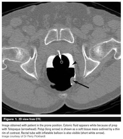
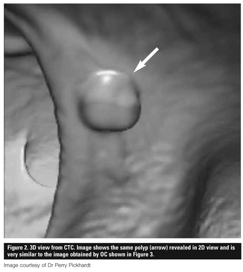
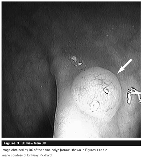
Radiologists will vary in their approach, based on their own experience and what equipment is available to them. The authors use the Viatronix workstation that Pickhardt used and a combination of 2D and 3D viewing, but rely on primary 3D interpretation. An example of a CTC study with a comparison colonoscopy image is shown in Figures 1, 2, and 3.
CT colonography versus standard optical colonoscopy
There is a steep learning curve when using either CTC or OC technology. For CTC, meticulous attention to technique, adequate time for careful analysis, and up-to-date equipment are crucial to the production of acceptable results. For OC, careful training and a dedication to absolute patient safety are needed to ensure accurate results and a safe and comfortable patient experience.
OC has very high sensitivity and specificity, making it the criterion standard for colorectal examination. In addition, OC images can often be used to distinguish between hyperplastic and adenomatous polyps on the basis of appearance or with the use of fluorescence imaging, and OC has the significant advantage of permitting immediate biopsy, polypectomy, or both if required.
The procedure is not perfect and lesions can be missed. Reports show miss rates of 26% to 27% for polyps less than 5 mm, 13% for polyps 6 to 9 mm, and 2% to 6% for polyps greater than 10 mm.[13,14] Overall OC is felt to have an accuracy of approximately 97%.
It does suffer from a less than 100% completion rate, although incompletion rates are very low in the hands of expert colonoscopists with the availability of double-balloon variable-stiffness adult and pediatric colonoscopes.
Risk of perforation is rare in experienced hands, ranging from 0.016%[15] to 0.22%.[16] Intravenous sedation is generally used for colonoscopy. Sedation complications are extremely rare. Most patients tolerate the procedure well and find the colon preparation to have been more of a challenge than the actual procedure. Colonoscopy has the additional advantage of zero radiation.
CTC does not require sedation, eliminating the need for an intravenous line, the potential side effects of sedation, and the need for monitoring. This allows patients to be released unaccompanied immediately following the procedure and to return to normal activity, including driving an automobile.
Comparing the level of patient comfort during the two examinations is difficult as OC patients are sedated and typically have little recollection of the study. Perforation rates during CTC are even lower than during OC.[16]
Inability to complete a CTC study is also rare, although a poorly distensible segment of colon may hamper CTC interpretation. Such segments may require endoscopic assessment.
An advantage of CTC over OC is its ability to detect significant extracolonic findings.[17] For example, CTC can identify renal cell carcinomas (4%), abdominal aortic aneurysms (5%), and lymphadenopathy (6%). The evaluation of solid organs in the abdomen is limited when compared with a standard contrast-enhanced CT scan because of the nonintravenous nature of contrast agents and low-dose radiation protocols used in CTC.
Studies comparing the accuracy of CTC and OC have previously shown widely varying results. Cotton,[15] an endoscopist, found a sensitivity of only 55% for polyps greater than 10 mm and 39% for polyps greater than 6 mm, whereas Pickhardt,[12] a radiologist, reported sensitivities of 93.8% and 88.7%, respectively, for polyps of the same size.
Disparities may be related to differences in experience and the quality of equipment used by these two research groups. In line with Pickhardt’s results, several recent large studies[16] have provided further strong evidence that CTC is capable of high accuracy.
Unpublished and preliminary results from the NIH-funded American College of Radiology Imaging Network (ACRIN) study were presented by Dr Johnson at this fall’s ACRIN meeting. The study involved 2531 patients in 15 US centres who had OC and CTC on the same day.
The sensitivity and specificity statistics from the study include a 90% per patient sensitivity for polyps greater than 10 mm—on par with OC sensitivity. This study also reported very high negative predictive value percentages, which are vital for an effective screening study.
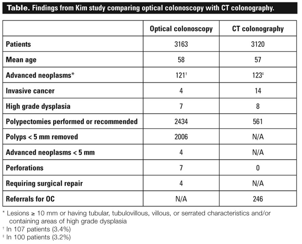
A second large study published recently by Kim[16] compared primary CTC in 3120 consecutive adults (mean age 57) with primary OC in 3163 consecutive adults (mean age 58). The results shown in the Table include the finding that only 246 CTC patients (7.9%) needed to be referred for OC.
Other relevant findings from this study include the fact that only 20 polyps less than 10 mm were histologically advanced in 15 patients (a rate of 0.2%). Also, only three patients with a total of four polyps less than 10 mm had high-grade dysplasia (a rate of 0.05%) and no subcentimetre cancers were found.
The authors concluded that “CTC and OC screening methods resulted in similar detection rates for advanced neoplasms within the same general population.” The primary CTC group underwent fewer optical colonoscopies and polypectomies, suggesting that CTC might be used effectively as a screening filter for therapeutic OC.
Perhaps such a combined approach would result in greater overall compliance for CRC screening.
Looking ahead
Of the imaging technologies used to view the entire colorectum, optical colonoscopy is still considered the criterion standard. However, when screening average-risk patients for CRC, CT colonography should be considered.
Earlier studies that showed poor sensitivity for CTC have now been largely refuted. Assuming there is state-of-the-art equipment and technical expertise in performing and interpreting the studies, CTC has high sensitivity.
The primary methodological issue that has prevented endoscopists from fully embracing CTC is based on the assumption that polyps less than 5 to 6 mm require biopsy or excision because advanced neoplasms may occasionally be small.
Nevertheless, the extremely low prevalence of advanced neoplasia or frank carcinoma in these small lesions may justify CTC management and permit following these patients with a repeat examination on a more frequent schedule and checking for interval increase in size.
As of March 2008, the American Cancer Society has added CTC to its list of acceptable front-line screening modalities.[18]
Future developments in CTC will include even more sophisticated and user-friendly workstation tools, which will reduce interpretation times and allow the incorporation of computer-aided diagnosis as a primary review to identify suspicious areas for the radiologist to reconcile.
Advances will also permit less vigorous colon cleansing.[19] Already some studies show promise for CTC with unprepared colons.[5] Advances on the horizon for OC include increasingly sophisticated light refraction systems that can identify areas of dysplasia not previously visible, wider angle lenses permitting greater view around folds to improve accuracy, instruments that will “walk” into the colon on their own motorized legs and negotiate colonic angulations more easily, and further improvements in analgesia to eliminate patient discomfort.
As well as improving the accuracy of both OC and CTC images, it is hoped that these developments will improve the currently poor screening rates in BC and Canada for a prevalent and deadly cancer.
Competing interests
Dr Flak receives fees for consultancy with Canadian Diagnostic Centres (BC), a private clinic that provides screening CT colonography. Dr Forster is the salaried medical director for Canada Diagnostic Centres, but neither he nor his professional practice group hold an equity position. Dr Pezim owns a clinic that undertakes colonoscopy examinations. Dr Pezim occasionally sends and receives referrals to and from Canada Diagnostic Services; in neither case is a fee exchanged.
References
1. National Cancer Institute of Canada. Review of trends in colorectal cancer. www.ncic.cancer.ca (accessed 16 March 2008).
2. British Columbia Vital Statistics Agency. Ministry of Health Planning. Death-related statistics. In: Selected vital statistics and health status indicators. 129th annual report, 2000. 130th annual report, 2001.
3. Rabeneck L, Paszat LF. A population-based estimate of the extent of colorectal cancer screening in Ontario. Am J Gastroenterol 2004;99:1141-1144.
4. McGregor SE, Hilsden RJ, Li FX, et al. Low uptake of colorectal cancer screening three years after release of national recommendations for screening. Am J Gastroenterol 2007;102:1736-1738.
5. Callstrom MR, Johnson CD, Fletcher JG, et al. CT colonography without cathartic preparation: Feasibility study. Radiology 2001;219:693-698.
6. Shinners TJ, Pickhardt PJ, Taylor AJ, et al. Patient-controlled room air insufflation versus automated carbon dioxide delivery for CT colonography. AJR Am J Roentgenol 2006;186:1491-1496.
7. Park SH, Ha HK, Kim MJ. False-negative results at multi-detector row CT colonography: Multivariate analysis of causes for missed lesions. Radiology 2005;235:495-502.
8. Morrin MM, Farrell RJ, Keogan MT, et al. CT colonography: Colonic distention improved by dual positioning but not intravenous glucagon. Eur Radiol 2002;12:525-530.
9. Macari M, Bini EJ, Xue X, et al. Colorectal neoplasms: Prospective comparison of thin-section low-dose multi-detector row CT colonography and conventional colonoscopy for detection. Radiology 2002;332:383-392.
10. Brenner DJ, Elliston CD. Estimated radiation risks potentially associated with full-body CT screening. Radiology 2004;232:735-738.
11. Brenner DJ. Estimating cancer risks from pediatric CT: Going from the qualitative to the quantitative. Pediatr Radiol 2002;32:228-231.
12. Pickhardt PJ, Choi JR, Hwang I, et al. Computed tomographic virtual colonoscopy to screen for colorectal neoplasia in asymptomatic adults. N Engl J Med 2003;349:2191-2200.
13. Rex DK, Cutler CS, Lemmel GT, et al. Colonoscopic miss rates of adenomas determined by back-to-back colonoscopies. Gastroenterology 1997;112:24-28.
14. van Rijn JC, Reitsma JB, Bossuyt PM, et al. Polyp miss rate determined by tandem colonoscopy: A systematic review. Am J Gastroenterol 2006;101:343-350.
15. Cotton PB, Durkalski VL, Pineau BC, et al. Computed tomographic colonography (virtual colonoscopy): A multicenter comparison with standard colonoscopy for detection of colorectal neoplasia. JAMA 2004;291:1713-1719.
16. Kim DH, Pickhardt PJ, Taylor AJ, et al. CT colonography versus colonoscopy for the detection of advanced neoplasia. N Engl J Med 2007;357:1403-1412.
17. Hellstrom M, Svensson MH, Lasson A. Extracolonic and incidental findings on CT colonography (virtual colonoscopy). AJR Am J Roentgenol 2004;182:631-638.
18. Health Groups Issue Updated Colorectal Cancer Screening Guidelines. www.cancer.org
19. Lefere P, Gryspeerdt S, Baekelandt M, et al. Laxative-free CT colonography. AJR Am J Roentgenol 2004;183:945-948.
Dr Flak provides professional services on a contract basis to Canadian Diagnostic Centres, where CT colonography studies are performed for screening purposes. Dr Forster is an associate professor and vice-chairman of research in the Department of Radiology at the University of British Columbia, and medical director of Canada Diagnostic Centres (BC). Dr Pezim is the medical director of the Pezim Clinic, a colorectal diagnostic centre in Vancouver, and a coauthor of The Intelligent Patient Guide to Colorectal Cancer.