
Cryptogenic multifocal ulcerous stenosing enteritis presenting as refractory iron deficiency anemia
Issue: BCMJ,
vol. 68, No. 8, October 2026
|
Pages 285-288 |
Clinical Case Reports | doi.org/10.66794/2c6a
ABSTRACT: Cryptogenic multifocal ulcerous stenosing enteritis is a rare idiopathic disorder characterized by recurrent superficial ulcerations and multiple strictures of the small intestine, typically occurring without systemic inflammation. We report the case of a 50-year-old man with a 20-year history of postprandial abdominal pain, iron deficiency anemia, and low albumin. Initial investigations were unrevealing, and the patient was treated with iron supplementation. With progressive symptoms, endoscopic evaluation suggested eosinophilic esophagitis and enteritis; however, he did not respond to conventional therapies, including systemic corticosteroids and dupilumab. Subsequent bidirectional endoscopy and magnetic resonance enterography were nondiagnostic. Capsule endoscopy later revealed diffuse multifocal ulcerative disease throughout the small intestine, with capsule retention at a suspected stricture. Double-balloon antegrade and retrograde enteroscopy was unsuccessful in identifying disease or retrieving the capsule. Ultimately, laparoscopic small bowel resection removed the affected segment and retrieved the retained capsule. Histopathologic examination demonstrated multifocal superficial ulcerations with associated strictures in the absence of granulomas or transmural inflammation, consistent with cryptogenic multifocal ulcerous stenosing enteritis. The patient experienced resolution of symptoms. This case illustrates the diagnostic challenges associated with cryptogenic multifocal ulcerous stenosing enteritis and underscores the importance of considering this rare entity in patients with unexplained chronic anemia and small bowel obstructive symptoms.
Cryptogenic multifocal ulcerous stenosing enteritis is rare and challenging to diagnose but should be considered in patients with unexplained chronic anemia and small bowel obstructive symptoms.
Cryptogenic multifocal ulcerous stenosing enteritis (CMUSE) is a rare inflammatory pathology of unknown origin, with multiple ulcerative strictures confined to the small bowel. With less than 100 published cases worldwide as of 2020,[1] CMUSE has probably been underdiagnosed or misdiagnosed, mainly as Crohn disease or NSAID-induced enteropathy.[2] This case report demonstrates the complexity of diagnosing CMUSE and the importance of an accurate diagnosis to guide treatment.
Case data
A 50-year-old man had experienced postprandial abdominal pain, iron deficiency anemia, and low albumin since he was in his mid-20s. His ethnicity was half Chinese and half Central European. His medical history was significant for exercise-induced asthma and numerous food allergies. Family history was negative for gastrointestinal disorders. He had initially come to medical attention in 2002 with shortness of breath on exertion, which was attributed to an exacerbation of his known exercise-induced asthma. Despite treatment for the latter, his dyspnea did not improve.
Subsequent blood work to investigate the cause of the patient’s dyspnea revealed microcytic anemia, with a hemoglobin level of 85 g/L and a ferritin level of 20 ng/mL. He had no use of NSAIDs, including both over-the-counter and prescription products. He did not use alcohol. At the time, he did not report abdominal symptoms. Bidirectional endoscopy, a barium swallow, and a barium enema did not reveal a cause of the marked anemia. He was advised to start oral iron supplementation, which helped normalize his hemoglobin.
Two years after the diagnosis of microcytic anemia, the patient developed intermittent postprandial left upper quadrant pain, bloating, nausea, and vomiting associated with partial small bowel obstructions. His weight was stable, with a low to normal BMI of 20; however, he was unable to gain weight despite a concentrated effort, consistent with malabsorption.
The diagnoses of eosinophilic esophagitis and eosinophilic enteritis were rendered based on biopsy findings from a repeat esophagogastroduodenoscopy in 2010. However, neither topical steroid nor proton pump inhibitor treatment improved the patient’s symptoms. He had taken oral iron supplements for decades to treat his chronic iron deficiency, presumed secondary to eosinophilic esophagitis and eosinophilic enteritis; however, in 2024, he required monthly IV iron infusions because his anemia had stopped responding to oral iron supplementation and his hemoglobin was hovering around 105 g/L. The monthly IV iron infusions did not increase his hemoglobin beyond 110 g/L, and his ferritin remained low, at 10 to 20 ng/mL.
A hematology consult excluded a primary hematological cause of the anemia. Concurrent with the decrease in hemoglobin, the patient’s episodes of postprandial left upper quadrant abdominal pain became more frequent and more severe, such that he opted for a trial of treatment in 2024. Because prior biopsy had suggested eosinophilic esophagitis and eosinophilic enteritis, he was trialed on oral budesonide for several months, then dupilumab for 6 months. Neither of these treatments improved his abdominal pain, and he remained anemic and hypoalbuminemic.
In late 2024, the patient underwent updated gastrointestinal investigations, including bidirectional endoscopy and magnetic resonance enterography (a previous MRI in 2021 showed no abnormalities). The only pertinent positive finding was chronic inactive gastritis and esophagitis without eosinophils on the upper endoscopy, but this did not account for his degree of anemia and hypoalbuminemia. Colonoscopy showed no abnormalities.
Given the ongoing suspicion of gastrointestinal-related blood loss, the patient had a capsule endoscopy in February 2025, which revealed diffuse multifocal ulcerative disease of the small bowel (jejunum) with spontaneous oozing from multiple ulcer sites. The capsule was retained at the site of involvement in a presumed stricture.
Double-balloon antegrade and retrograde enteroscopy in June 2025 did not show macroscopic or microscopic abnormalities. The capsule was not located, because it was beyond the reach of the double-balloon studies.
In September 2025, the patient’s diagnosis was still uncertain. A laparoscopic small bowel resection was performed to remove the diseased section (165 cm of strictures and shallow ulcers [Figure 1]) and retrieve the capsule. Pathology revealed multifocal superficial ulcerations and strictures, negative for granulomas and transmural inflammation. There were reactive lymph nodes with abundant hemosiderin-laden macrophages [Figure 2]. This identified the diagnosis of CMUSE.


One month postoperatively, the patient’s hemoglobin and albumin had normalized, yet his ferritin remained low, at 20 ng/mL. His postprandial abdominal pain had not returned since his bowel resection. Capsule endoscopy was chosen as the preferred method for surveillance going forward, because it was the only modality of investigation that had been able to identify the disease process in this patient. He was scheduled for repeat capsule endoscopy 6 months postoperatively.
Discussion
Diagnosis of CMUSE is based on history, clinical features, CT or magnetic resonance enterography, small intestinal endoscopy (capsule endoscopy or double-balloon enteroscopy), and histology of the small bowel.[2-4]
Chronic or relapsing subileus episodes resulting from multiple small intestinal strictures are a leading clinical symptom, along with chronic iron deficiency anemia from small bowel occult blood loss, which may precede the diagnosis for several years.[2] There may be fatigue, edema, or growth retardation.[5]
Blood analysis tends to show iron deficiency anemia and low albumin.[6] Inflammatory markers and other biological signs of systemic inflammation are usually not abnormal.[5,6]
CT or magnetic resonance enterography may show short annular ulcerative strictures at the jejunum and/or ileum, while the inflammatory ulcerative strictures can also be recognized as multiple intestinal foci on fluorodeoxyglucose PET-CT.[3,6]
Capsule endoscopy reveals intestinal ulcerations; however, there is an increased risk of capsule retention due to the presence of strictures.[6] In British Columbia, capsule endoscopy can be ordered by a gastroenterologist if gastrointestinal bleeding in the small intestine is suspected, but only after other investigations have ruled out other causes (as per the Medical Services Commission Payment Schedule). Antegrade or retrograde enteroscopy can confirm the presence of one or more short ulcerative strictures with normal mucosa between;[6] however, sometimes the involved area cannot be reached, as in the case of our patient.
Histological findings include superficial ulceration, fibrosis and nonspecific inflammation of the mucosa and submucosa, and vascular changes without transmural inflammation.[4]
The Table summarizes the differential diagnoses of CMUSE.[2,6-9] Crohn disease is an important differential diagnosis, with skip lesions and cobblestoning on macroscopic appearance, while histology reveals transmural inflammation, and the typical lesions are longitudinal and fissured ulcers, which are prone to fistula formation and intestinal perforation.[4,5] NSAID-induced enteropathy is very similar to CMUSE on endoscopy and histology. In NSAID-induced enteropathy, there tends to be gradual resolution of disease with NSAID cessation.[5] Genetic testing for PLA2G4A mutations, genes associated with prostaglandin metabolism, can also distinguish these conditions.[6] Other differential diagnoses include intestinal tuberculosis, vasculitis, chronic ulcerative jejunitis, collagen sprue, autoimmune or eosinophilic enteritis, and chronic nonspecific ulcers of the small intestine.[2,5,6]
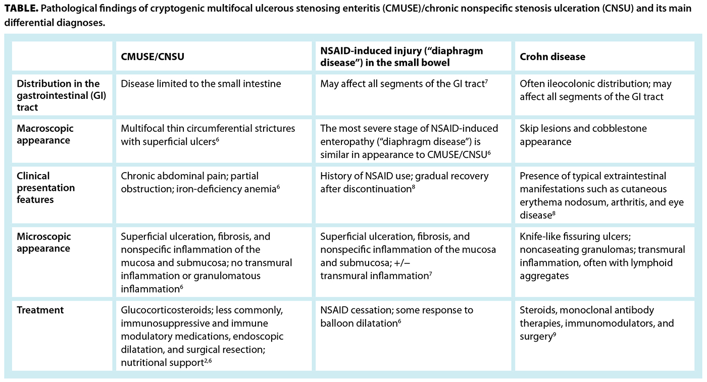
Chronic nonspecific stenosing ulceration is a similar entity that affects predominantly Japanese patients and is associated with loss-of-function mutations in the SLCO2A1 gene.[6]
Medical treatment consists of systemic and local glucocorticosteroids at individual-specific doses;[2,6] however, most patients develop corticosteroid dependence,[2] and some will relapse or develop steroid resistance.[5,6] There have been case reports of long-term remission using immunosuppressive and immune modulatory medications typically used in Crohn disease and ulcerative colitis, such as mesalazine, mercaptopurine, azathioprine, thalidomide, and infliximab.[6]
Endoscopic balloon dilatation can alleviate symptoms and prevent small bowel resection.[2,5] Surgical resection is a last resort, reserved for those with small bowel obstruction or patients who do not respond to medical treatment.[2,6] However, patients can easily relapse, and surgery poses a risk of complications such as short bowel syndrome.[2,4]
Furthermore, supportive care by means of enteral or even parenteral nutrition and iron and vitamin supplements may improve symptoms and promote mucosal healing.[5,6]
Summary
In this case, the patient had received systemic steroids as treatment for eosinophilic esophagitis and eosinophilic enteritis, which did not promote remission of CMUSE. Ultimately, a small bowel resection was performed to alleviate his symptoms and clarify his diagnosis. Continued follow-up is necessary to monitor for relapse. This case highlights the complexity of diagnosing CMUSE and the importance of an accurate diagnosis to guide treatment.
Competing interests
None declared.
This article has been peer reviewed.

This work is licensed under a Creative Commons Attribution-NonCommercial-NoDerivatives 4.0 International License.
References
1. Woodall CB, Raines D. S4842: CMUSE in the United States: A case series. Am J Gastroenterol 2024;119(10S):S3054-S3055. https://doi.org/10.14309/01.ajg.0001048736.39719.9e.
2. Kohoutová D, Bártová J, Tachecí I, et al. Cryptogenic multifocal ulcerous stenosing enteritis: A review of the literature. Gastroenterol Res Pract 2013;2013:918031. https://doi.org/10.1155/2013/918031.
3. Zhang X, Ma L, Xiao M, et al. Imaging diagnosis of cryptogenic multifocal ulcerous stenosing enteritis. Insights Imaging 2025;16:55. https://doi.org/10.1186/s13244-025-01931-9.
4. Lew C, Jain A, Chua J, Wong A. Cryptogenic multifocal ulcerous stenosing enteritis: A difficult diagnosis. ANZ J Surg 2022;92:2373-2375. https://doi.org/10.1111/ans.17489.
5. Chang C, Jiang C, Miao Y, et al. A case report of intestinal obstruction caused by cryptogenic multifocal ulcerous stenosing enteritis. BMC Gastroenterol 2020;20:300. https://doi.org/10.1186/s12876-020-01450-5.
6. Moreels TG, Singh A. Updates on the diagnosis and management of cryptogenic multifocal ulcerative stenosing enteropathy (CMUSE) and non-steroidal enteropathy. Best Pract Res Clin Gastroenterol 2023;64-65:101847. https://doi.org/10.1016/j.bpg.2023.101847.
7. Chung SH, Jo Y, Ryu SR, et al. Diaphragm disease compared with cryptogenic multifocal ulcerous stenosing enteritis. World J Gastroenterol 2011;17:2873-2876. https://doi.org/10.3748/wjg.v17.i23.2873.
8. Hudson L, Grayson N, Toomey D. Cryptogenic multifocal ulcerative stenosing enteritis (CMUSE): A rare case and a review of the literature. J Surg Case Rep 2025;2026:rjaf977. https://doi.org/10.1093/jscr/rjaf977.
9. Cushing K, Higgins PDR. Management of Crohn disease: A review. JAMA 2021;325:69-80. https://doi:10.1001/jama.2020.18936.
Dr Chan is a clinical associate professor in the Department of Family Practice at the University of British Columbia and a locum at various family practices across the Lower Mainland and the Fraser Valley. Dr Yang is an associate professor in the Department of Pathology and Laboratory Medicine at UBC and a staff anatomical pathologist at St. Paul’s Hospital. Dr Villanyi is a clinical assistant professor in the Division of Geriatric Medicine at UBC and a staff geriatrician at Vancouver General Hospital and UBC Hospital. Dr Lee is an associate professor in the Department of Pathology and Laboratory Medicine at UBC and a staff anatomical pathologist at St. Paul’s Hospital.