Anatomy in a distributed model of medical education
The move to a distributed model of medical education presented significant challenges to the way we train students in gross human anatomy and in histology. To meet these challenges, we took advantage of some innovative technology that allowed instruction to occur simultaneously at the three UBC medical program sites.
Innovative technology has allowed students at all three UBC medical school sites to have the same lectures, laboratories, and examinations, and to access micrographs and digitized slides when receiving their training in gross human anatomy and histology.
The move to a distributed model of medical education in BC presented significant challenges when determining how to train students in gross human anatomy and in histology. These challenges have been met with the help of technology that allows us to provide instruction simultaneously at the three University of British Columbia MD undergraduate program sites in Victoria, Prince George, and Vancouver.
Gross human anatomy
Training in anatomy has been integrated into the medical curriculum during the first 2 years of study, and in the new distributed education model it occurs at all three sites. Among the major challenges we faced was to provide students with equivalent experiences (lectures, laboratories, and examinations) at all three sites and to ensure students and faculty members at the distributed sites were fully integrated with those at UBC.
In the distributed gross anatomy program, dissection is the cornerstone of the educational experience; however, dissections are streamlined to focus on major and clinically relevant structures and difficult dissections are replaced by, or augmented with, plastinated prosections. A group of six students is assigned to each cadaver, and cadavers are moved to the distributed sites when students move to these locations.
During the first 3 months of the program, when all students are at the main UBC campus, key concepts related to the general organization of the nervous system are established and students focus on learning the anatomy of the body’s trunk (thorax, abdomen, pelvis, and perineum). When students move to the three distributed sites, material learned during the first 3 months is reviewed and new material related to the anatomy of the upper limb, lower limb, head, and neck is learned.
The gross anatomy program is coordinated by a director at UBC. At each of the distributed sites, local faculty and staff oversee the program and provide content expertise. Faculty and staff at all three sites communicate regularly to ensure the program runs smoothly and students have equivalent experiences. New lecture halls and dissection laboratories have been constructed at all three sites.
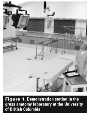
Each site has the capacity to transmit lectures and laboratory demonstrations to the other two sites (see Figure 1). This ensures faculty members at the University of Northern British Columbia (UNBC) and the University of Victoria (UVic) have the opportunity to deliver sessions simultaneously to the entire class located at all three sites, and that faculty can travel to any site to deliver material to the entire class.
Most gross anatomy sessions begin with a lecture followed by a 2- to 3- hour laboratory. Each laboratory begins with a pre-lab talk in which the faculty member responsible for the session demonstrates how the dissection is to be done and reviews the major features to be identified.
These pre-lab talks and demonstrations are transmitted to multiple speakers and monitors in the laboratories at all three sites simultaneously. Following the orientation sessions, students perform the dissections and identify key structures listed in handouts that are the same at all sites.
Laboratory examinations are set by the program director at UBC in consultation with the faculty at UNBC and UVic. Equivalent examinations are set up at all three sites and are taken by students on the same day.
The body donation program is administered at UBC. Cadavers are moved by funeral homes to laboratories at UNBC and UVic in December and then are returned to UBC for cremation the following year.
Histology
With the expanded class size, histology has been integrated into various blocks of the first 2 years of the medical and dental programs. Histology instruction now consists of 21 lectures and 34 2-hour laboratory sessions spread over 2 years, and combines computer-assisted instruction, conventional microscope use, conventional instruction, and the study of virtual slides.
In the first term of the first year, all students attend sessions in Vancouver. In the following three terms, all lectures and laboratory sessions are delivered by videoconference simultaneously at the three sites.
A new histology web site, linked to an online curriculum system for all medical students, has been created to accommodate distributed learning. It includes all pertinent learning materials: lecture and laboratory handouts; hundreds of light and electron micrographs; interactive quizzes; and a “virtual slide” box with hundreds of digitized slides.
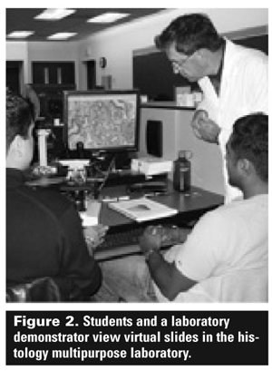
The virtual slides are digitized variable magnification images of the histology slide collection. Students have access to the web site 24 hours a day, 7 days a week, either from their computer workstations in the new histology multipurpose laboratory (see Figure 2) or from their personal computers. Students also have the opportunity to use conventional microscopes and glass slides stored in lockers at each workstation.
The multipurpose laboratory at UBC has been designed to accommodate 320 students. Each of the 40 workstations has four computers and monitors. Two students share each monitor in the laboratory. This approach facilitates interactive, self-directed learning and fosters team building. Similar but smaller labs with a virtually identical design have been constructed at UVic and UNBC.
Each laboratory session provides many opportunities for scientific discovery. Students can reach conclusions on their own, share observations with peers, and discuss them with instructors. A strength of the teaching program is the active participation by laboratory instructors and teaching assistants at all three sites.
Each laboratory session begins with a prerecorded lab talk broadcast to all three sites, and ends with a short self-assessment quiz in the form of a PowerPoint presentation. Four end-of-term laboratory examinations are delivered by videoconference using a large screen at each of the three sites.
Several years into using a distributed model to deliver our problem-based learning medical curriculum, we find the gross anatomy and histology programs have both operated with surprisingly few problems.
Competing interests
None declared.
Drs Vogl and Ovalle are professors in the Department of Cellular and Physiological Sciences, Faculty of Medicine, UBC.